7F5A | pdb_00007f5a

DNQX-bound GluK2-2xNeto2 complex


Experimental Data Snapshot

  • Method: ELECTRON MICROSCOPY
  • Resolution: 6.40 Å
  • Aggregation State: PARTICLE 
  • Reconstruction Method: SINGLE PARTICLE 

wwPDB Validation 3D Report Full Report

Validation slider image for 7F5A

This is version 1.2 of the entry. See complete history

Literature

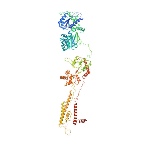
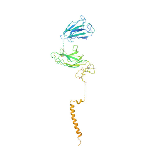
Kainate receptor modulation by NETO2.

He, L.Sun, J.Gao, Y.Li, B.Wang, Y.Dong, Y.An, W.Li, H.Yang, B.Ge, Y.Zhang, X.C.Shi, Y.S.Zhao, Y.

(2021) Nature 599: 325-329

  • DOI: https://doi.org/10.1038/s41586-021-03936-y
  • Primary Citation Related Structures: 
    7F56, 7F57, 7F59, 7F5A, 7F5B

  • PubMed Abstract: 

    Glutamate-gated kainate receptors are ubiquitous in the central nervous system of vertebrates, mediate synaptic transmission at the postsynapse and modulate transmitter release at the presynapse 1-7 . In the brain, the trafficking, gating kinetics and pharmacology of kainate receptors are tightly regulated by neuropilin and tolloid-like (NETO) proteins 8-11 . Here we report cryo-electron microscopy structures of homotetrameric GluK2 in complex with NETO2 at inhibited and desensitized states, illustrating variable stoichiometry of GluK2-NETO2 complexes, with one or two NETO2 subunits associating with GluK2. We find that NETO2 accesses only two broad faces of kainate receptors, intermolecularly crosslinking the lower lobe of ATD A/C , the upper lobe of LBD B/D and the lower lobe of LBD A/C , illustrating how NETO2 regulates receptor-gating kinetics. The transmembrane helix of NETO2 is positioned proximal to the selectivity filter and competes with the amphiphilic H1 helix after M4 for interaction with an intracellular cap domain formed by the M1-M2 linkers of the receptor, revealing how rectification is regulated by NETO2.


  • Organizational Affiliation
    • National Laboratory of Biomacromolecules, CAS Center for Excellence in Biomacromolecules, Institute of Biophysics, Chinese Academy of Sciences, Beijing, China.

Macromolecule Content 

  • Total Structure Weight: 533.29 kDa 
  • Atom Count: 31,178 
  • Modeled Residue Count: 3,912 
  • Deposited Residue Count: 4,682 
  • Unique protein chains: 2

Macromolecules

Find similar proteins by:|  3D Structure
Entity ID: 1
MoleculeChains  Sequence LengthOrganismDetailsImage
Glutamate receptor ionotropic, kainate 2
A, B, C, D
908Rattus norvegicusMutation(s): 3 
Gene Names: Grik2Glur6
Membrane Entity: Yes 
UniProt
Find proteins for P42260 (Rattus norvegicus)
Explore P42260 
Go to UniProtKB:  P42260
Entity Groups
Sequence Clusters30% Identity50% Identity70% Identity90% Identity95% Identity100% Identity
UniProt GroupP42260
Glycosylation
Glycosylation Sites: 1Go to GlyGen: P42260-1
Sequence Annotations
Expand
Reference Sequence
Find similar proteins by:|  3D Structure
Entity ID: 2
MoleculeChains  Sequence LengthOrganismDetailsImage
Neuropilin and tolloid-like protein 2
E, F
525Rattus norvegicusMutation(s): 0 
Gene Names: Neto2Btcl2
Membrane Entity: Yes 
UniProt
Find proteins for C6K2K4 (Rattus norvegicus)
Explore C6K2K4 
Go to UniProtKB:  C6K2K4
Entity Groups
Sequence Clusters30% Identity50% Identity70% Identity90% Identity95% Identity100% Identity
UniProt GroupC6K2K4
Sequence Annotations
Expand
Reference Sequence

Oligosaccharides

Help  
Entity ID: 3
MoleculeChains Length2D Diagram GlycosylationD Interactions
beta-D-mannopyranose-(1-4)-2-acetamido-2-deoxy-beta-D-glucopyranose-(1-4)-2-acetamido-2-deoxy-beta-D-glucopyranose
G, H, I, J
3N-Glycosylation
Glycosylation Resources
GlyTouCan: G15407YE
GlyCosmos: G15407YE
GlyGen: G15407YE

Small Molecules

Ligands 1 Unique
IDChains Name / Formula / InChI Key2D Diagram3D Interactions
NAG

Query on NAG


Download:Ideal Coordinates CCD File
K [auth A]
L [auth A]
M [auth B]
N [auth B]
O [auth B]
K [auth A],
L [auth A],
M [auth B],
N [auth B],
O [auth B],
P [auth B],
Q [auth C],
R [auth D],
S [auth D],
T [auth D]
2-acetamido-2-deoxy-beta-D-glucopyranose
C8 H15 N O6
OVRNDRQMDRJTHS-FMDGEEDCSA-N

Experimental Data & Validation

Experimental Data

  • Method: ELECTRON MICROSCOPY
  • Resolution: 6.40 Å
  • Aggregation State: PARTICLE 
  • Reconstruction Method: SINGLE PARTICLE 

Structure Validation

View Full Validation Report


Entry History 

& Funding Information

Deposition Data


Funding OrganizationLocationGrant Number
Chinese Academy of SciencesChinaXDB37030304

Revision History  (Full details and data files)

  • Version 1.0: 2021-09-29
    Type: Initial release
  • Version 1.1: 2021-12-01
    Changes: Database references
  • Version 1.2: 2024-11-06
    Changes: Data collection, Structure summary