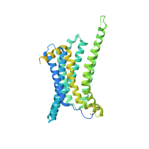
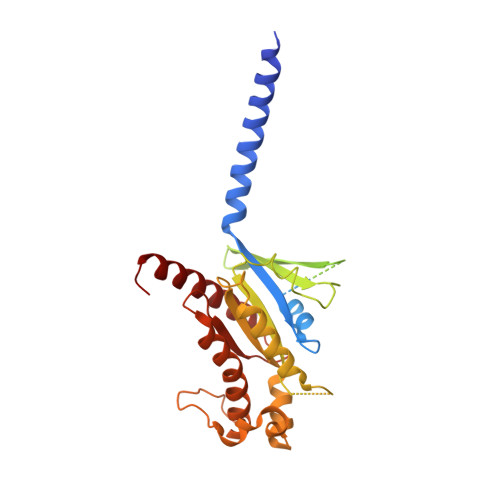
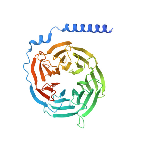
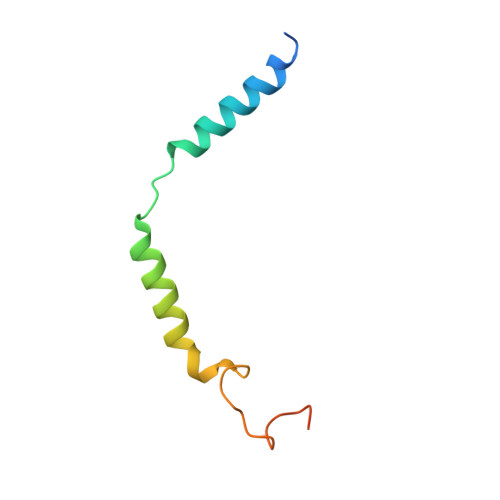
Melanocortin-4 receptor (MC4R) plays a central role in the regulation of energy homeostasis. Its high sequence similarity to other MC receptor family members, low agonist selectivity and the lack of structural information concerning MC4R-specific activation have hampered the development of MC4R-seletive therapeutics to treat obesity. Here, we report four high-resolution structures of full-length MC4R in complex with the heterotrimeric G s protein stimulated by the endogenous peptide ligand α-MSH, FDA-approved drugs afamelanotide (Scenesse™) and bremelanotide (Vyleesi™), and a selective small-molecule ligand THIQ, respectively. Together with pharmacological studies, our results reveal the conserved binding mode of peptidic agonists, the distinctive molecular details of small-molecule agonist recognition underlying receptor subtype selectivity, and a distinct activation mechanism for MC4R, thereby offering new insights into G protein coupling. Our work may facilitate the discovery of selective therapeutic agents targeting MC4R.
Organizational Affiliation:
Department of Biophysics and Department of Pathology of Sir Run Run Shaw Hospital, Zhejiang University School of Medicine, Hangzhou, Zhejiang, China.
Liangzhu Laboratory, Zhejiang University Medical Center, Hangzhou, Zhejiang, China.
MOE Frontier Science Center for Brain Research and Brain-Machine Integration, Zhejiang University School of Medicine, Hangzhou, Zhejiang, China.
Key Laboratory of Immunity and Inflammatory Diseases of Zhejiang Province, Hangzhou, Zhejiang, China.
The CAS Key Laboratory of Receptor Research, Shanghai Institute of Materia Medica, Chinese Academy of Sciences, Shanghai, China.
The National Center for Drug Screening, Shanghai Institute of Materia Medica, Chinese Academy of Sciences, Shanghai, China.
University of Chinese Academy of Sciences, Beijing, China.
School of Pharmacy, Fudan University, Shanghai, China.
Department of Cell Biology and Cancer Institute of the Second Affiliated Hospital, Zhejiang University School of Medicine, Hangzhou, China; Institute of Gastroenterology, Zhejiang University, Hangzhou, China; Collaborative Innovation Center for Diagnosis and Treatment of Infectious Diseases, Hangzhou, Zhejiang, China; Department of Molecular Genetics, University of Toronto, Toronto, Ontario, Canada.
Department of Physiology and Pathophysiology, School of Basic Medical Sciences, Peking University; Key Laboratory of Molecular Cardiovascular Science, Ministry of Education, Beijing, China.
Key Laboratory Experimental Teratology of the Ministry of Education, Department of Biochemistry and Molecular Biology, School of Basic Medical Sciences, Cheeloo College of Medicine, Shandong University, Jinan, Shandong, China.
Department of Microbiology and The Children's Hospital, Zhejiang University School of Medicine, Hangzhou, Zhejiang, China.
Innovation Institute for Artificial Intelligence in Medicine of Zhejiang University, College of Pharmaceutical Sciences, Zhejiang University, Hangzhou, Zhejiang, China.
The CAS Key Laboratory of Receptor Research, Shanghai Institute of Materia Medica, Chinese Academy of Sciences, Shanghai, China. mwwang@simm.ac.cn.
The National Center for Drug Screening, Shanghai Institute of Materia Medica, Chinese Academy of Sciences, Shanghai, China. mwwang@simm.ac.cn.
University of Chinese Academy of Sciences, Beijing, China. mwwang@simm.ac.cn.
School of Pharmacy, Fudan University, Shanghai, China. mwwang@simm.ac.cn.
School of Life Science and Technology, ShanghaiTech University, Shanghai, China. mwwang@simm.ac.cn.
Department of Pharmacology, Fudan University, Shanghai, China. mwwang@simm.ac.cn.
Department of Biophysics and Department of Pathology of Sir Run Run Shaw Hospital, Zhejiang University School of Medicine, Hangzhou, Zhejiang, China. zhang_yan@zju.edu.cn.
Liangzhu Laboratory, Zhejiang University Medical Center, Hangzhou, Zhejiang, China. zhang_yan@zju.edu.cn.
MOE Frontier Science Center for Brain Research and Brain-Machine Integration, Zhejiang University School of Medicine, Hangzhou, Zhejiang, China. zhang_yan@zju.edu.cn.
Key Laboratory of Immunity and Inflammatory Diseases of Zhejiang Province, Hangzhou, Zhejiang, China. zhang_yan@zju.edu.cn.