


Structural basis for MTA1c-mediated DNA N6-adenine methylation
Chen, J., Hu, R., Chen, Y., Lin, X., Xiang, W., Chen, H., Yao, C., Liu, L.(2022) Nat Commun 13: 3257
Experimental Data Snapshot
Starting Model: experimental
View more details
Entity ID: 1 | |||||
|---|---|---|---|---|---|
| Molecule | Chains | Sequence Length | Organism | Details | Image |
| Transmembrane protein, putative | A [auth B] | 142 | Tetrahymena thermophila SB210 | Mutation(s): 0 Gene Names: TTHERM_00439330 | 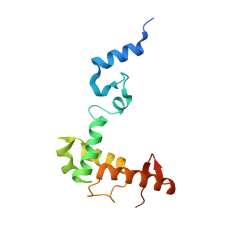 |
UniProt | |||||
Entity Groups | |||||
| Sequence Clusters | 30% Identity50% Identity70% Identity90% Identity95% Identity100% Identity | ||||
| UniProt Group | I7M8B9 | ||||
Sequence AnnotationsExpand | |||||
Reference Sequence | |||||
Entity ID: 2 | |||||
|---|---|---|---|---|---|
| Molecule | Chains | Sequence Length | Organism | Details | Image |
| MT-a70 family protein | B [auth A] | 247 | Tetrahymena thermophila SB210 | Mutation(s): 0 Gene Names: MTA1 EC: 2.1.1.348 | 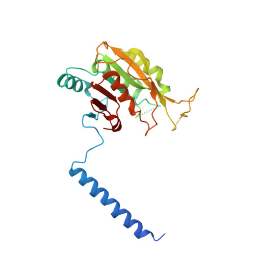 |
UniProt | |||||
Entity Groups | |||||
| Sequence Clusters | 30% Identity50% Identity70% Identity90% Identity95% Identity100% Identity | ||||
| UniProt Group | Q22GC0 | ||||
Sequence AnnotationsExpand | |||||
Reference Sequence | |||||
| Ligands 1 Unique | |||||
|---|---|---|---|---|---|
| ID | Chains | Name / Formula / InChI Key | 2D Diagram | 3D Interactions | |
| SAM (Subject of Investigation/LOI) Download:Ideal Coordinates CCD File | C [auth A] | S-ADENOSYLMETHIONINE C15 H22 N6 O5 S MEFKEPWMEQBLKI-FCKMPRQPSA-N |  | ||
| Length ( Å ) | Angle ( ˚ ) |
|---|---|
| a = 137.032 | α = 90 |
| b = 137.032 | β = 90 |
| c = 61.629 | γ = 120 |
| Software Name | Purpose |
|---|---|
| PHENIX | refinement |
| PHENIX | refinement |
| HKL-3000 | data reduction |
| HKL-3000 | data scaling |
| PHASER | phasing |
| Funding Organization | Location | Grant Number |
|---|---|---|
| National Natural Science Foundation of China (NSFC) | China | 32022047 |


