7F3N | pdb_00007f3n

Structure of PopP2 in apo form


Experimental Data Snapshot

  • Method: X-RAY DIFFRACTION
  • Resolution: 2.35 Å
  • R-Value Free: 
    0.231 (Depositor), 0.233 (DCC) 
  • R-Value Work: 
    0.193 (Depositor), 0.208 (DCC) 
  • R-Value Observed: 
    0.197 (Depositor) 

Starting Model: experimental
View more details

wwPDB Validation 3D Report Full Report

Validation slider image for 7F3N

This is version 1.1 of the entry. See complete history

Literature

Secondary-structure switch regulates the substrate binding of a YopJ family acetyltransferase.

Xia, Y.Zou, R.Escouboue, M.Zhong, L.Zhu, C.Pouzet, C.Wu, X.Wang, Y.Lv, G.Zhou, H.Sun, P.Ding, K.Deslandes, L.Yuan, S.Zhang, Z.M.

(2021) Nat Commun 12: 5969-5969

  • DOI: https://doi.org/10.1038/s41467-021-26183-1
  • Primary Citation Related Structures: 
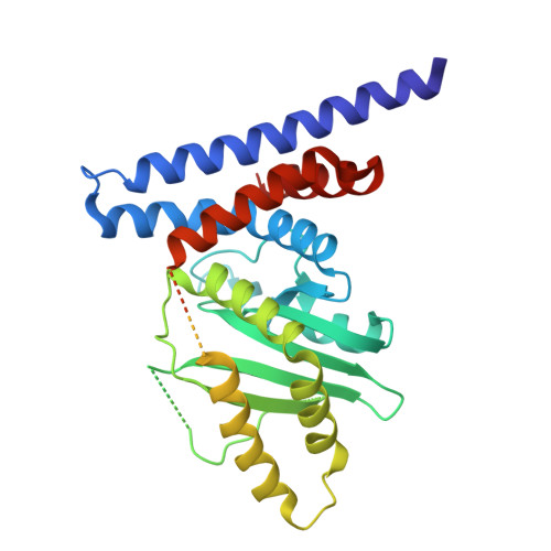
    7F3N

  • PubMed Abstract: 

    The Yersinia outer protein J (YopJ) family effectors are widely deployed through the type III secretion system by both plant and animal pathogens. As non-canonical acetyltransferases, the enzymatic activities of YopJ family effectors are allosterically activated by the eukaryote-specific ligand inositol hexaphosphate (InsP6). However, the underpinning molecular mechanism remains undefined. Here we present the crystal structure of apo-PopP2, a YopJ family member secreted by the plant pathogen Ralstonia solanacearum. Structural comparison of apo-PopP2 with the InsP6-bound PopP2 reveals a substantial conformational readjustment centered in the substrate-binding site. Combining biochemical and computational analyses, we further identify a mechanism by which the association of InsP6 with PopP2 induces an α-helix-to-β-strand transition in the catalytic core, resulting in stabilization of the substrate recognition helix in the target protein binding site. Together, our study uncovers the molecular basis governing InsP6-mediated allosteric regulation of YopJ family acetyltransferases and further expands the paradigm of fold-switching proteins.


  • Organizational Affiliation
    • International Cooperative Laboratory of Traditional Chinese Medicine Modernization and Innovative Drug Development of Chinese Ministry of Education (MOE), College of Pharmacy, Jinan University, 510632, Guangzhou, China.

Macromolecule Content 

  • Total Structure Weight: 37.74 kDa 
  • Atom Count: 1,887 
  • Modeled Residue Count: 247 
  • Deposited Residue Count: 343 
  • Unique protein chains: 1

Macromolecules

Find similar proteins by:|  3D Structure
Entity ID: 1
MoleculeChains  Sequence LengthOrganismDetailsImage
Type III effector protein popp2343Ralstonia pseudosolanacearum GMI1000Mutation(s): 0 
Gene Names: popP2RSc0868
UniProt
Find proteins for Q8Y125 (Ralstonia nicotianae (strain ATCC BAA-1114 / GMI1000))
Explore Q8Y125 
Go to UniProtKB:  Q8Y125
Entity Groups
Sequence Clusters30% Identity50% Identity70% Identity90% Identity95% Identity100% Identity
UniProt GroupQ8Y125
Sequence Annotations
Expand
Reference Sequence

Experimental Data & Validation

Experimental Data

  • Method: X-RAY DIFFRACTION
  • Resolution: 2.35 Å
  • R-Value Free:  0.231 (Depositor), 0.233 (DCC) 
  • R-Value Work:  0.193 (Depositor), 0.208 (DCC) 
  • R-Value Observed: 0.197 (Depositor) 
Space Group: P 32 2 1
Unit Cell:
Length ( Å )Angle ( ˚ )
a = 89.36α = 90
b = 89.36β = 90
c = 79.871γ = 120
Software Package:
Software NamePurpose
PHENIXrefinement
HKL-2000data scaling
Cootmodel building
HKL-2000data scaling
PHASERphasing

Structure Validation

View Full Validation Report


Entry History 

Deposition Data

Revision History  (Full details and data files)

  • Version 1.0: 2021-11-17
    Type: Initial release
  • Version 1.1: 2023-11-29
    Changes: Data collection, Refinement description