7F21

L-lactate oxidase with D-lactate


Experimental Data Snapshot

  • Method: X-RAY DIFFRACTION
  • Resolution: 1.38 Å
  • R-Value Free: 0.224 
  • R-Value Work: 0.196 
  • R-Value Observed: 0.197 

wwPDB Validation   3D Report Full Report


Ligand Structure Quality Assessment 


This is version 2.1 of the entry. See complete history


Literature

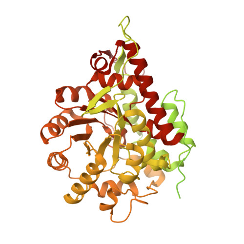
Dynamic interactions in the l-lactate oxidase active site facilitate substrate binding at pH4.5.

Furubayashi, N.Inaka, K.Kamo, M.Umena, Y.Matsuoka, T.Morimoto, Y.

(2021) Biochem Biophys Res Commun 568: 131-135

  • DOI: https://doi.org/10.1016/j.bbrc.2021.06.078
  • Primary Citation of Related Structures:  
    7F1Y, 7F20, 7F21, 7F22

  • PubMed Abstract: 

    The crystal structure of l-lactate oxidase in complex with l-lactate was solved at a 1.33 Å resolution. The electron density of the bound l-lactate was clearly shown and comparisons of the free form and substrate bound complexes demonstrated that l-lactate was bound to the FMN and an additional active site within the enzyme complex. l-lactate interacted with the related side chains, which play an important role in enzymatic catalysis and especially the coupled movement of H265 and D174, which may be essential to activity. These observations not only reveal the enzymatic mechanism for l-lactate binding but also demonstrate the dynamic motion of these enzyme structures in response to substrate binding and enzymatic reaction progression.


  • Organizational Affiliation

    MARUWA Foods and Biosciences, Inc., 170-1, Tsutsui-cho, Yamatokoriyama, Nara, 639-1123, Japan.


Macromolecules
Find similar proteins by:  (by identity cutoff)  |  3D Structure
Entity ID: 1
MoleculeChains Sequence LengthOrganismDetailsImage
L-lactate oxidase
A, B
720Aerococcus viridansMutation(s): 0 
UniProt
Find proteins for Q44467 (Aerococcus viridans (strain ATCC 11563 / DSM 20340 / CCUG 4311 / JCM 20461 / NBRC 12219 / NCTC 8251 / M1))
Explore Q44467 
Go to UniProtKB:  Q44467
Entity Groups  
Sequence Clusters30% Identity50% Identity70% Identity90% Identity95% Identity100% Identity
UniProt GroupQ44467
Sequence Annotations
Expand
  • Reference Sequence
Experimental Data & Validation

Experimental Data

  • Method: X-RAY DIFFRACTION
  • Resolution: 1.38 Å
  • R-Value Free: 0.224 
  • R-Value Work: 0.196 
  • R-Value Observed: 0.197 
  • Space Group: I 4
Unit Cell:
Length ( Å )Angle ( ˚ )
a = 132.781α = 90
b = 132.781β = 90
c = 92.07γ = 90
Software Package:
Software NamePurpose
REFMACrefinement
XDSdata reduction
Aimlessdata scaling
MOLREPphasing

Structure Validation

View Full Validation Report


Ligand Structure Quality Assessment 


Entry History 

Deposition Data

Revision History  (Full details and data files)

  • Version 1.0: 2022-03-23
    Type: Initial release
  • Version 2.0: 2023-11-15
    Changes: Atomic model, Data collection
  • Version 2.1: 2023-11-29
    Changes: Refinement description