7F0H | pdb_00007f0h

Structural and functional characterization of bovine G1P[5] rotavirus VP8* protein


Experimental Data Snapshot

  • Method: X-RAY DIFFRACTION
  • Resolution: 1.70 Å
  • R-Value Free: 
    0.188 (Depositor), 0.188 (DCC) 
  • R-Value Work: 
    0.167 (Depositor), 0.167 (DCC) 
  • R-Value Observed: 
    0.168 (Depositor) 

Starting Model: experimental
View more details

wwPDB Validation 3D Report Full Report

Validation slider image for 7F0H

This is version 1.1 of the entry. See complete history

Literature

Structural and functional characterization of bovine G1P[5] rotavirus VP8* protein.

Dang, L.Su, Y.Qi, J.Wu, Z.Li, D.Wang, M.Zhang, Q.Wang, H.Bai, R.Duan, Z.Sun, X.

(2021) Virology 563: 116-125

  • DOI: https://doi.org/10.1016/j.virol.2021.08.009
  • Primary Citation Related Structures: 
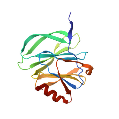
    7F0H

  • PubMed Abstract: 

    The widely used rotavirus (RV) vaccine, Rotateq, contained reassortment strains of human and bovine G1/2/3/4P[5] RVs. The functional and structural features of bovine G1P[5] VP8* were investigated. Bovine G1P[5] VP8* was identified to interact with sialic acids and sialic acid-containing glycans. In addition, P[5] VP8* recognized α-Gal histo-blood group antigens (HBGAs). Bovine G1P[5] VP8* did not hemagglutinate the tested red blood cells. The crystal structure of P[5] VP8* was determined at 1.7 Å. Structural superimposition revealed that P[5] VP8* was most close to human P[8] VP8*, while much further to VP8*s of porcine P[7] and rhesus P[3]. Sequence alignment showed that amino acids of the putative glycan binding site in P[5] VP8* were different to those in P[3]/P[7] VP8*s, indicating that P[5] VP8* may interact with glycans using different mechanism. This study provided more understanding of P[5] RV infection and the interactions of RV VP8* and glycans.


  • Organizational Affiliation
    • National Health Commission Key Laboratory for Medical Virology and Viral Diseases, Beijing, 102206, China; National Institute for Viral Disease Control and Prevention, China CDC, Beijing, 102206, China; Inner Mongolia Hospital of Traditional Chinese Medicine, Hohhot, 010059, China.

Macromolecule Content 

  • Total Structure Weight: 36.17 kDa 
  • Atom Count: 3,032 
  • Modeled Residue Count: 322 
  • Deposited Residue Count: 322 
  • Unique protein chains: 1

Macromolecules

Find similar proteins by:|  3D Structure
Entity ID: 1
MoleculeChains  Sequence LengthOrganismDetailsImage
Outer capsid protein VP8*
A, B
161Rotavirus AMutation(s): 0 
UniProt
Find proteins for Q8JNB4 (Rotavirus A (strain RVA/Cow/United States/WC3/1981/G6P7[5]))
Explore Q8JNB4 
Go to UniProtKB:  Q8JNB4
Entity Groups
Sequence Clusters30% Identity50% Identity70% Identity90% Identity95% Identity100% Identity
UniProt GroupQ8JNB4
Sequence Annotations
Expand
Reference Sequence

Experimental Data & Validation

Experimental Data

  • Method: X-RAY DIFFRACTION
  • Resolution: 1.70 Å
  • R-Value Free:  0.188 (Depositor), 0.188 (DCC) 
  • R-Value Work:  0.167 (Depositor), 0.167 (DCC) 
  • R-Value Observed: 0.168 (Depositor) 
Space Group: I 21 21 21
Unit Cell:
Length ( Å )Angle ( ˚ )
a = 97.753α = 90
b = 102.364β = 90
c = 137.722γ = 90
Software Package:
Software NamePurpose
HKL-2000data scaling
PHENIXrefinement
PDB_EXTRACTdata extraction
HKL-2000data reduction
PHASERphasing

Structure Validation

View Full Validation Report


Entry History 

& Funding Information

Deposition Data


Funding OrganizationLocationGrant Number
National Natural Science Foundation of China (NSFC)China21934005

Revision History  (Full details and data files)

  • Version 1.0: 2021-12-29
    Type: Initial release
  • Version 1.1: 2023-11-29
    Changes: Data collection, Refinement description