


Crystal structure of arginine kinase (AK3) from the ciliate Paramecium tetraurelia
Otsuka, Y., Yokota, J., Yano, D., Uda, K., Suzuki, T., Sugiyama, S.To be published.
Experimental Data Snapshot
Starting Model: experimental
View more details
wwPDB Validation 3D Report Full Report

Entity ID: 1 | |||||
|---|---|---|---|---|---|
| Molecule | Chains | Sequence Length | Organism | Details | Image |
| arginine kinase | 401 | Paramecium tetraurelia | Mutation(s): 0 | 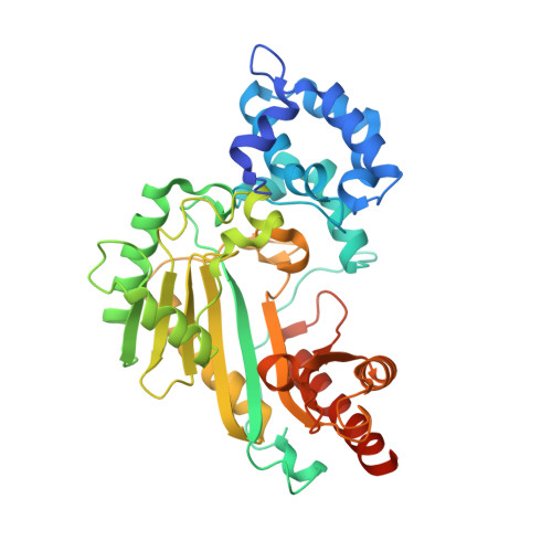 | |
UniProt | |||||
Entity Groups | |||||
| Sequence Clusters | 30% Identity50% Identity70% Identity90% Identity95% Identity100% Identity | ||||
| UniProt Group | A0C7I4 | ||||
Sequence AnnotationsExpand | |||||
Reference Sequence | |||||
| Length ( Å ) | Angle ( ˚ ) |
|---|---|
| a = 70.109 | α = 90 |
| b = 77.727 | β = 90 |
| c = 162.197 | γ = 90 |
| Software Name | Purpose |
|---|---|
| REFMAC | refinement |
| XDS | data reduction |
| Aimless | data scaling |
| REFMAC | phasing |
| Funding Organization | Location | Grant Number |
|---|---|---|
| Japan Science and Technology | Japan | JPMJTM19DC |
| Japan Society for the Promotion of Science (JSPS) | Japan | 19K06588 |
| Other private | Japan | 09-003-005 |


