Chasing the structural diversity of the transcription regulator Mycobacterium tuberculosis HigA2.
Richardson, W., Kang, G.W., Lee, H.J., Kwon, K.M., Kim, S., Kim, H.J.(2021) IUCrJ 8: 823-832
- PubMed: 34584743 Search on PubMedSearch on PubMed Central
- DOI: https://doi.org/10.1107/S2052252521007715
- Primary Citation Related Structures:
7EWC, 7EWD, 7EWE - PubMed Abstract:
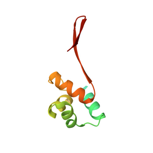
Transcription factors are the primary regulators of gene expression and recognize specific DNA sequences under diverse physiological conditions. Although they are vital for many important cellular processes, it remains unclear when and how transcription factors and DNA interact. The antitoxin from a toxin-antitoxin system is an example of negative transcriptional autoregulation: during expression of the cognate toxin it is suppressed through binding to a specific DNA sequence. In the present study, the antitoxin HigA2 from Mycobacterium tuberculosis M37Rv was structurally examined. The crystal structure of M. tuberculosis HigA2 comprises three sections: an N-terminal autocleavage region, an α-helix bundle which contains an HTH motif, and a C-terminal β-lid. The N-terminal region is responsible for toxin binding, but was shown to cleave spontaneously in its absence. The HTH motif performs a key role in DNA binding, with the C-terminal β-lid influencing the interaction by mediating the distance between the motifs. However, M. tuberculosis HigA2 exhibits a unique coordination of the HTH motif and no DNA-binding activity is detected. Three crystal structures of M. tuberculosis HigA2 show a flexible alignment of the HTH motif, which implies that the motif undergoes structural rearrangement to interact with DNA. This study reveals the molecular mechanisms of how transcription factors interact with partner proteins or DNA.
- Centre for Biomolecular Sciences, School of Pharmacy, University of Nottingham, Nottingham, United Kingdom.
Organizational Affiliation: