7EUY | pdb_00007euy

Crystal structure of the Lon-like protease MtaLonC with D582A mutation in complex with substrate polypeptide


Experimental Data Snapshot

  • Method: X-RAY DIFFRACTION
  • Resolution: 2.20 Å
  • R-Value Free: 
    0.211 (Depositor), 0.221 (DCC) 
  • R-Value Work: 
    0.182 (Depositor), 0.190 (DCC) 
  • R-Value Observed: 
    0.184 (Depositor) 

Starting Model: experimental
View more details

wwPDB Validation 3D Report Full Report

Validation slider image for 7EUY

This is version 1.1 of the entry. See complete history

Literature

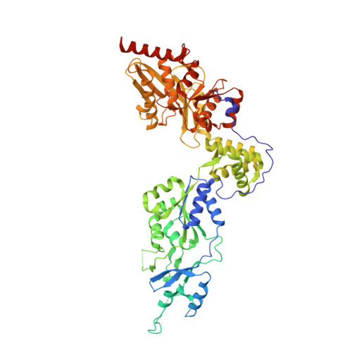
Processive cleavage of substrate at individual proteolytic active sites of the Lon protease complex.

Li, S.Hsieh, K.Y.Kuo, C.I.Su, S.C.Huang, K.F.Zhang, K.Chang, C.I.

(2021) Sci Adv 7: eabj9537-eabj9537

  • DOI: https://doi.org/10.1126/sciadv.abj9537
  • Primary Citation Related Structures: 
    7EUX, 7EUY, 7EV4, 7EV6, 7FID, 7FIE, 7FIZ

  • PubMed Abstract: 

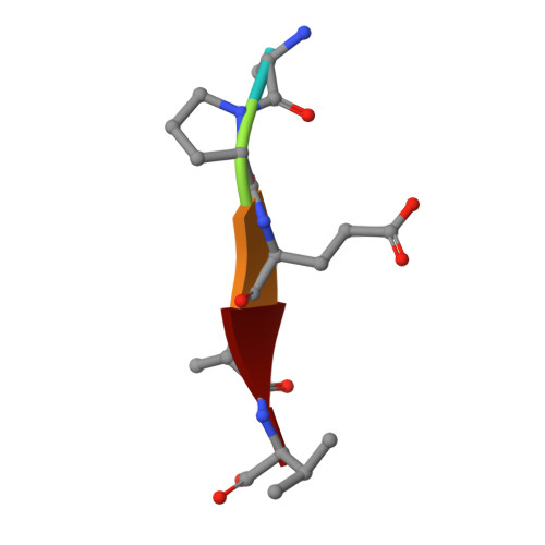
    The Lon protease is the prototype of a family of proteolytic machines with adenosine triphosphatase modules built into a substrate degradation chamber. Lon is known to degrade protein substrates in a processive fashion, cutting a protein chain processively into small peptides before commencing cleavages of another protein chain. Here, we present structural and biochemical evidence demonstrating that processive substrate degradation occurs at each of the six proteolytic active sites of Lon, which forms a deep groove that partially encloses the substrate polypeptide chain by accommodating only the unprimed residues and permits processive cleavage in the C-to-N direction. We identify a universally conserved acidic residue at the exit side of the binding groove indispensable for the proteolytic activity. This noncatalytic residue likely promotes processive proteolysis by carboxyl-carboxylate interactions with cleaved intermediates. Together, these results uncover a previously unrecognized mechanism for processive substrate degradation by the Lon protease.


  • Organizational Affiliation
    • MOE Key Laboratory for Membraneless Organelles and Cellular Dynamics, Hefei National Laboratory for Physical Sciences at the Microscale and Division of Life Sciences and Medicine, University of Science and Technology of China, Hefei 230027, China.

Macromolecule Content 

  • Total Structure Weight: 81.12 kDa 
  • Atom Count: 4,829 
  • Modeled Residue Count: 592 
  • Deposited Residue Count: 737 
  • Unique protein chains: 2

Macromolecules

Find similar proteins by:|  3D Structure
Entity ID: 1
MoleculeChains  Sequence LengthOrganismDetailsImage
Endopeptidase La732Meiothermus taiwanensisMutation(s): 1 
EC: 3.4.21.53
UniProt
Find proteins for C9DRU9 (Meiothermus taiwanensis)
Explore C9DRU9 
Go to UniProtKB:  C9DRU9
Entity Groups
Sequence Clusters30% Identity50% Identity70% Identity90% Identity95% Identity100% Identity
UniProt GroupC9DRU9
Sequence Annotations
Expand
Reference Sequence
Find similar proteins by:  Sequence   |   3D Structure  
Entity ID: 2
MoleculeChains  Sequence LengthOrganismDetailsImage
ALA-PRO-GLU-ALA-VALB [auth S]5Escherichia coliMutation(s): 0 

Small Molecules

Ligands 1 Unique
IDChains Name / Formula / InChI Key2D Diagram3D Interactions
PO4

Query on PO4


Download:Ideal Coordinates CCD File
C [auth A]PHOSPHATE ION
O4 P
NBIIXXVUZAFLBC-UHFFFAOYSA-K

Experimental Data & Validation

Experimental Data

  • Method: X-RAY DIFFRACTION
  • Resolution: 2.20 Å
  • R-Value Free:  0.211 (Depositor), 0.221 (DCC) 
  • R-Value Work:  0.182 (Depositor), 0.190 (DCC) 
  • R-Value Observed: 0.184 (Depositor) 
Space Group: P 6
Unit Cell:
Length ( Å )Angle ( ˚ )
a = 115.838α = 90
b = 115.838β = 90
c = 135.455γ = 120
Software Package:
Software NamePurpose
REFMACrefinement
PDB_EXTRACTdata extraction
HKL-2000data reduction
HKL-2000data scaling
PHASERphasing

Structure Validation

View Full Validation Report


Entry History 

& Funding Information

Deposition Data


Funding OrganizationLocationGrant Number
Ministry of Science and Technology (MoST, Taiwan)Taiwan108-2320-B-001-011-MY3
Academia Sinica (Taiwan)Taiwan--

Revision History  (Full details and data files)

  • Version 1.0: 2021-11-24
    Type: Initial release
  • Version 1.1: 2023-11-29
    Changes: Data collection, Refinement description