7EUN

Crystal structure of N(omega)-hydroxy-L-arginine hydrolase in complex with ABH


Experimental Data Snapshot

  • Method: X-RAY DIFFRACTION
  • Resolution: 1.28 Å
  • R-Value Free: 0.138 
  • R-Value Work: 0.115 
  • R-Value Observed: 0.116 

wwPDB Validation   3D Report Full Report


Ligand Structure Quality Assessment 


This is version 1.3 of the entry. See complete history


Literature

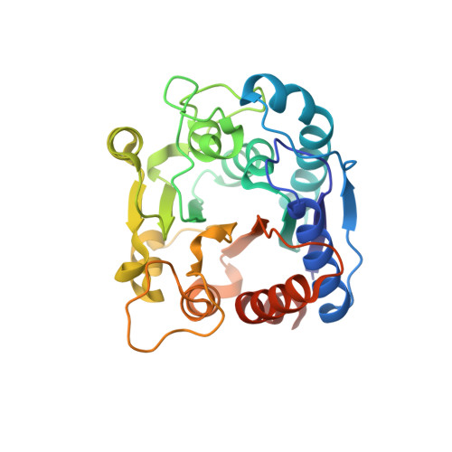
Catalytic mechanism of DcsB: Arginase framework used for hydrolyzing its inhibitor.

Oda, K.Sakaguchi, T.Matoba, Y.

(2022) Protein Sci 31: e4338-e4338

  • DOI: https://doi.org/10.1002/pro.4338
  • Primary Citation of Related Structures:  
    7EUK, 7EUL, 7EUN, 7EUQ

  • PubMed Abstract: 

    DcsB, an enzyme produced from the d-cycloserine biosynthetic gene cluster, displays moderate similarity to arginase in the sequence and three-dimensional structure. Arginase is a ubiquitous enzyme hydrolyzing l-arginine to generate l-ornithine and urea, whereas DcsB hydrolyzes N ω -hydroxy-l-arginine (l-NOHA), an arginase inhibitor, to generate l-ornithine and hydroxyurea. We determined the crystal structure of DcsB associated with l-ornithine and that with the tetrahedral derivative of 2(S)-amino-6-boronohexanoic acid, whose boron atom forms a covalent bond with an oxygen atom bridging two manganese ions at the active center. The substrate-binding pocket of DcsB is narrower than that of arginase, suggesting that DcsB is unsuitable for the binding of l-NOHA in an inhibitory manner. The transition state-like structure demonstrated that Asp210 and Glu241 have a role to trap a positively charged ion near the dimanganese cluster. Kinetic analysis using the mutated DcsB showed that the enzyme employs different catalytic mechanisms under the neutral and alkaline pH conditions. Glu241 in DcsB is likely involved in the recognition of the hydroxyguanidino group of l-NOHA, whereas Asp210, in cooperation with Glu241, seems to contribute to the reactivity toward the protonated l-NOHA, which is a preferable species under the neutral pH conditions. After entering of the protonated l-NOHA to the substrate-binding pocket of DcsB, a hydronium ion may be trapped at the positive ion-binding site. Then, the ion serves as a specific acid catalyst to facilitate the collapse of the tetrahedral intermediate of l-NOHA.


  • Organizational Affiliation

    Department of Virology, Institute of Biomedical and Health Sciences, Hiroshima University, Hiroshima, Japan.


Macromolecules
Find similar proteins by:  (by identity cutoff)  |  3D Structure
Entity ID: 1
MoleculeChains Sequence LengthOrganismDetailsImage
N(omega)-hydroxy-L-arginine amidinohydrolase
A, B
281Streptomyces lavendulaeMutation(s): 0 
Gene Names: dcsB
EC: 3.5.3.25
UniProt
Find proteins for D2Z025 (Streptomyces lavendulae)
Explore D2Z025 
Go to UniProtKB:  D2Z025
Entity Groups  
Sequence Clusters30% Identity50% Identity70% Identity90% Identity95% Identity100% Identity
UniProt GroupD2Z025
Sequence Annotations
Expand
  • Reference Sequence
Small Molecules
Ligands 3 Unique
IDChains Name / Formula / InChI Key2D Diagram3D Interactions
ABH (Subject of Investigation/LOI)
Query on ABH

Download Ideal Coordinates CCD File 
J [auth B]2(S)-AMINO-6-BORONOHEXANOIC ACID
C6 H15 B N O5
BLVGFZFOWWBCCZ-YFKPBYRVSA-N
MN (Subject of Investigation/LOI)
Query on MN

Download Ideal Coordinates CCD File 
C [auth A],
D [auth A],
F [auth B],
G [auth B]
MANGANESE (II) ION
Mn
WAEMQWOKJMHJLA-UHFFFAOYSA-N
MG (Subject of Investigation/LOI)
Query on MG

Download Ideal Coordinates CCD File 
E [auth A],
H [auth B],
I [auth B]
MAGNESIUM ION
Mg
JLVVSXFLKOJNIY-UHFFFAOYSA-N
Experimental Data & Validation

Experimental Data

  • Method: X-RAY DIFFRACTION
  • Resolution: 1.28 Å
  • R-Value Free: 0.138 
  • R-Value Work: 0.115 
  • R-Value Observed: 0.116 
  • Space Group: P 1
Unit Cell:
Length ( Å )Angle ( ˚ )
a = 46.577α = 83.54
b = 47.208β = 84.44
c = 59.641γ = 70.2
Software Package:
Software NamePurpose
PHENIXrefinement
Aimlessdata scaling
PDB_EXTRACTdata extraction
XDSdata reduction
PHENIXphasing

Structure Validation

View Full Validation Report


Ligand Structure Quality Assessment 


Entry History & Funding Information

Deposition Data


Funding OrganizationLocationGrant Number
Not funded--

Revision History  (Full details and data files)

  • Version 1.0: 2022-05-18
    Type: Initial release
  • Version 1.1: 2022-06-15
    Changes: Database references
  • Version 1.2: 2022-06-22
    Changes: Database references
  • Version 1.3: 2023-11-29
    Changes: Data collection, Refinement description