7ESI

Crystal structure of the collagenase unit of a Vibrio collagenase from Vibrio harveyi VHJR7 at 1. 8 angstrom resolution.


Experimental Data Snapshot

  • Method: X-RAY DIFFRACTION
  • Resolution: 1.80 Å
  • R-Value Free: 0.187 
  • R-Value Work: 0.159 
  • R-Value Observed: 0.160 

wwPDB Validation   3D Report Full Report


This is version 1.1 of the entry. See complete history


Literature

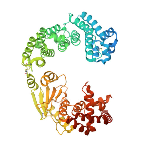
Structure of Vibrio collagenase VhaC provides insight into the mechanism of bacterial collagenolysis.

Wang, Y.Wang, P.Cao, H.Y.Ding, H.T.Su, H.N.Liu, S.C.Liu, G.Zhang, X.Li, C.Y.Peng, M.Li, F.Li, S.Chen, Y.Chen, X.L.Zhang, Y.Z.

(2022) Nat Commun 13: 566-566

  • DOI: https://doi.org/10.1038/s41467-022-28264-1
  • Primary Citation of Related Structures:  
    7ESI

  • PubMed Abstract: 

    The collagenases of Vibrio species, many of which are pathogens, have been regarded as an important virulence factor. However, there is little information on the structure and collagenolytic mechanism of Vibrio collagenase. Here, we report the crystal structure of the collagenase module (CM) of Vibrio collagenase VhaC and the conformation of VhaC in solution. Structural and biochemical analyses and molecular dynamics studies reveal that triple-helical collagen is initially recognized by the activator domain, followed by subsequent cleavage by the peptidase domain along with the closing movement of CM. This is different from the peptidolytic mode or the proposed collagenolysis of Clostridium collagenase. We propose a model for the integrated collagenolytic mechanism of VhaC, integrating the functions of VhaC accessory domains and its collagen degradation pattern. This study provides insight into the mechanism of bacterial collagenolysis and helps in structure-based drug design targeting of the Vibrio collagenase.


  • Organizational Affiliation

    State Key Laboratory of Microbial Technology, Shandong University, Qingdao, 266237, China.


Macromolecules
Find similar proteins by:  (by identity cutoff)  |  3D Structure
Entity ID: 1
MoleculeChains Sequence LengthOrganismDetailsImage
Collagenase unit (CU)613Vibrio harveyi VHJR7Mutation(s): 0 
UniProt
Find proteins for A0A8B3DGT3 (Vibrio harveyi)
Explore A0A8B3DGT3 
Go to UniProtKB:  A0A8B3DGT3
Entity Groups  
Sequence Clusters30% Identity50% Identity70% Identity90% Identity95% Identity100% Identity
UniProt GroupA0A8B3DGT3
Sequence Annotations
Expand
  • Reference Sequence

Find similar proteins by:  Sequence   |   3D Structure  

Entity ID: 2
MoleculeChains Sequence LengthOrganismDetailsImage
Peptide P114Vibrio harveyi VHJR7Mutation(s): 0 
Entity Groups  
Sequence Clusters30% Identity50% Identity70% Identity90% Identity95% Identity100% Identity
Sequence Annotations
Expand
  • Reference Sequence

Find similar proteins by:  Sequence   |   3D Structure  

Entity ID: 3
MoleculeChains Sequence LengthOrganismDetailsImage
Peptide P212Vibrio harveyi VHJR7Mutation(s): 0 
Entity Groups  
Sequence Clusters30% Identity50% Identity70% Identity90% Identity95% Identity100% Identity
Sequence Annotations
Expand
  • Reference Sequence
Experimental Data & Validation

Experimental Data

  • Method: X-RAY DIFFRACTION
  • Resolution: 1.80 Å
  • R-Value Free: 0.187 
  • R-Value Work: 0.159 
  • R-Value Observed: 0.160 
  • Space Group: P 1 21 1
Unit Cell:
Length ( Å )Angle ( ˚ )
a = 74.444α = 90
b = 50.068β = 104.72
c = 90.681γ = 90
Software Package:
Software NamePurpose
PHENIXrefinement
PDB_EXTRACTdata extraction
HKL-2000data reduction
HKL-2000data scaling
AutoSolphasing

Structure Validation

View Full Validation Report


Entry History 

Deposition Data

Revision History  (Full details and data files)

  • Version 1.0: 2022-02-09
    Type: Initial release
  • Version 1.1: 2023-02-22
    Changes: Database references