7EQ9

Cryo-EM structure of designed protein nanoparticle TIP60 (Truncated Icosahedral Protein composed of 60-mer fusion proteins)


Experimental Data Snapshot

  • Method: ELECTRON MICROSCOPY
  • Resolution: 3.30 Å
  • Aggregation State: PARTICLE 
  • Reconstruction Method: SINGLE PARTICLE 

wwPDB Validation   3D Report Full Report


This is version 1.2 of the entry. See complete history


Literature

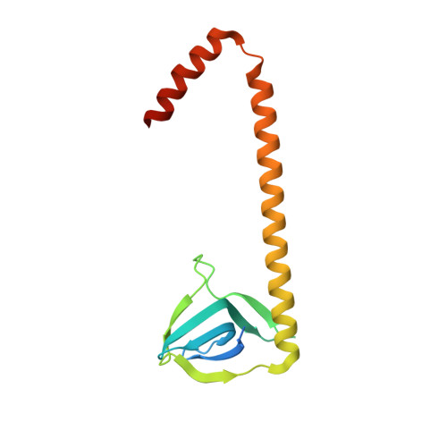
Icosahedral 60-meric porous structure of designed supramolecular protein nanoparticle TIP60.

Obata, J.Kawakami, N.Tsutsumi, A.Nasu, E.Miyamoto, K.Kikkawa, M.Arai, R.

(2021) Chem Commun (Camb) 57: 10226-10229

  • DOI: https://doi.org/10.1039/d1cc03114g
  • Primary Citation of Related Structures:  
    7EQ9

  • PubMed Abstract: 

    Supramolecular protein nanoparticles and nanocages have potential in a broad range of applications. Recently, we developed a uniform supramolecular protein nanoparticle, TIP60, symmmetrically self-assembled from fusion proteins of a pentameric Sm-like protein and a dimeric MyoX-coil domain. Herein, we report the icosahedral 60-meric structure of TIP60 solved using single-particle cryo-electron microscopy. Interestingly, the structure revealed 20 regular-triangle-like pores on the surface. TIP60 and its mutants have many modifiable sites on their exterior and interior surfaces. The TIP60 architecture will be useful in the development of biomedical and biochemical nanoparticles/nanocages for future applications.


  • Organizational Affiliation

    Department of Biomolecular Innovation, Institute for Biomedical Sciences, Interdisciplinary Cluster for Cutting Edge Research, Shinshu University, Ueda, Nagano 386-8567, Japan. rarai@shinshu-u.ac.jp.


Macromolecules
Find similar proteins by:  (by identity cutoff)  |  3D Structure
Entity ID: 1
MoleculeChains Sequence LengthOrganismDetailsImage
TIP60153synthetic constructMutation(s): 0 
Entity Groups  
Sequence Clusters30% Identity50% Identity70% Identity90% Identity95% Identity100% Identity
Sequence Annotations
Expand
  • Reference Sequence
Experimental Data & Validation

Experimental Data

  • Method: ELECTRON MICROSCOPY
  • Resolution: 3.30 Å
  • Aggregation State: PARTICLE 
  • Reconstruction Method: SINGLE PARTICLE 
EM Software:
TaskSoftware PackageVersion
RECONSTRUCTIONRELION3.1
MODEL REFINEMENTPHENIX1.19

Structure Validation

View Full Validation Report


Entry History & Funding Information

Deposition Data


Funding OrganizationLocationGrant Number
Japan Society for the Promotion of Science (JSPS)JapanJP19H02522
Japan Society for the Promotion of Science (JSPS)JapanJP18K05324
Japan Society for the Promotion of Science (JSPS)JapanJP17KK0104
Japan Society for the Promotion of Science (JSPS)JapanJP16K05841
Japan Agency for Medical Research and Development (AMED)JapanJP20am0101115

Revision History  (Full details and data files)

  • Version 1.0: 2021-09-15
    Type: Initial release
  • Version 1.1: 2022-03-16
    Changes: Database references
  • Version 1.2: 2023-11-29
    Changes: Data collection, Refinement description