7EMS

Crystal Structure of the HasAp L85A Mutant Capturing Iron Tetraphenylporphyrin


Experimental Data Snapshot

  • Method: X-RAY DIFFRACTION
  • Resolution: 1.85 Å
  • R-Value Free: 0.204 
  • R-Value Work: 0.167 
  • R-Value Observed: 0.169 

wwPDB Validation   3D Report Full Report


Ligand Structure Quality Assessment 


This is version 1.2 of the entry. See complete history


Literature

Tetraphenylporphyrin Enters the Ring: First Example of a Complex between Highly Bulky Porphyrins and a Protein.

Shisaka, Y.Sakakibara, E.Suzuki, K.Stanfield, J.K.Onoda, H.Ueda, G.Hatano, M.Sugimoto, H.Shoji, O.

(2022) Chembiochem 23: e202200095-e202200095

  • DOI: https://doi.org/10.1002/cbic.202200095
  • Primary Citation of Related Structures:  
    7EMO, 7EMP, 7EMQ, 7EMR, 7EMS, 7EMT, 7EMU, 7EMV, 7EMW, 7VM1

  • PubMed Abstract: 

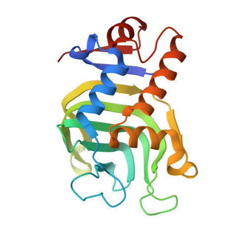
    Tetraphenylporphyrin (TPP) is a symmetrically substituted synthetic porphyrin whose properties can be readily modified, providing it with significant advantages over naturally occurring porphyrins. Herein, we report the first example of a stable complex between a native biomolecule, the haemoprotein HasA, and TPP as well as its derivatives. The X-ray crystal structures of nine different HasA-TPP complexes were solved at high resolutions. HasA capturing TPP derivatives was also demonstrated to inhibit growth of the opportunistic pathogen Pseudomonas aeruginosa. Mutant variants of HasA binding FeTPP were shown to possess a different mode of coordination, permitting the cyclopropanation of styrene.


  • Organizational Affiliation

    Department of Chemistry, Graduate School of Science, Nagoya University, Furo-cho, Chikusa-ku, Nagoya, 464-8602, Japan.


Macromolecules
Find similar proteins by:  (by identity cutoff)  |  3D Structure
Entity ID: 1
MoleculeChains Sequence LengthOrganismDetailsImage
Heme acquisition protein HasAp
A, B, C
185Pseudomonas aeruginosa PAO1Mutation(s): 1 
Gene Names: hasApPA3407
UniProt
Find proteins for G3XD33 (Pseudomonas aeruginosa (strain ATCC 15692 / DSM 22644 / CIP 104116 / JCM 14847 / LMG 12228 / 1C / PRS 101 / PAO1))
Explore G3XD33 
Go to UniProtKB:  G3XD33
Entity Groups  
Sequence Clusters30% Identity50% Identity70% Identity90% Identity95% Identity100% Identity
UniProt GroupG3XD33
Sequence Annotations
Expand
  • Reference Sequence
Experimental Data & Validation

Experimental Data

  • Method: X-RAY DIFFRACTION
  • Resolution: 1.85 Å
  • R-Value Free: 0.204 
  • R-Value Work: 0.167 
  • R-Value Observed: 0.169 
  • Space Group: P 1 21 1
Unit Cell:
Length ( Å )Angle ( ˚ )
a = 31.119α = 90
b = 125.104β = 99.45
c = 66.176γ = 90
Software Package:
Software NamePurpose
XDSdata reduction
Aimlessdata scaling
REFMACrefinement
PDB_EXTRACTdata extraction
MOLREPphasing

Structure Validation

View Full Validation Report


Ligand Structure Quality Assessment 


Entry History & Funding Information

Deposition Data


Funding OrganizationLocationGrant Number
Japan Society for the Promotion of Science (JSPS)JapanJP18H02084
Japan Science and TechnologyJapanJPMJCR15P3
Japan Society for the Promotion of Science (JSPS)JapanJP18J15250

Revision History  (Full details and data files)

  • Version 1.0: 2022-04-20
    Type: Initial release
  • Version 1.1: 2022-08-17
    Changes: Database references
  • Version 1.2: 2023-11-29
    Changes: Data collection, Refinement description