Color-Tuning Mechanism of the Lit Form of Orange Carotenoid Protein.
Han, M.H., Yang, H.W., Yoon, J., Villafani, Y., Song, J.Y., Pan, C.H., Park, K., Cho, Y., Song, J.J., Kim, S.J., Park, Y.I., Park, J.(2023) Mol Cells 46: 513-525
- PubMed: 37587751 Search on PubMedSearch on PubMed Central
- DOI: https://doi.org/10.14348/molcells.2023.2186
- Primary Citation Related Structures:
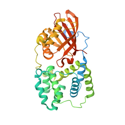
7EKR - PubMed Abstract:
Orange carotenoid protein (OCP) of photosynthetic cyanobacteria binds to ketocarotenoids noncovalently and absorbs excess light to protect the host organism from light-induced oxidative damage. Herein, we found that mutating valine 40 in the α3 helix of Gloeocapsa sp. PCC 7513 (G l OCP1) resulted in blue- or red-shifts of 6-20 nm in the absorption maxima of the lit forms. We analyzed the origins of absorption maxima shifts by integrating X-ray crystallography, homology modeling, molecular dynamics simulations, and hybrid quantum mechanics/molecular mechanics calculations. Our analysis suggested that the single residue mutations alter the polar environment surrounding the bound canthaxanthin, thereby modulating the degree of charge transfer in the photoexcited state of the chromophore. Our integrated investigations reveal the mechanism of color adaptation specific to OCPs and suggest a design principle for color-specific photoswitches.
- Department of Physics, Korea Advanced Institute of Science and Technology (KAIST), Daejeon 34141, Korea.
Organizational Affiliation: