7EFN

Crystal structure of the gastric proton pump K791S/E820D/Y340N/E936V in (BYK)E2BeF state


Experimental Data Snapshot

  • Method: X-RAY DIFFRACTION
  • Resolution: 3.20 Å
  • R-Value Free: 0.308 
  • R-Value Work: 0.268 
  • R-Value Observed: 0.270 

wwPDB Validation   3D Report Full Report


Ligand Structure Quality Assessment 


This is version 1.2 of the entry. See complete history


Literature

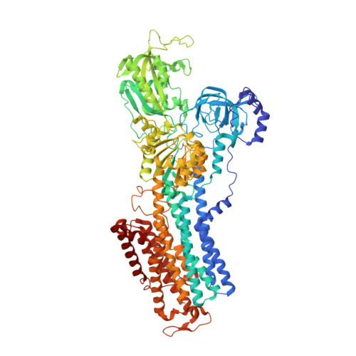
Gastric proton pump with two occluded K + engineered with sodium pump-mimetic mutations.

Abe, K.Yamamoto, K.Irie, K.Nishizawa, T.Oshima, A.

(2021) Nat Commun 12: 5709-5709

  • DOI: https://doi.org/10.1038/s41467-021-26024-1
  • Primary Citation of Related Structures:  
    7EFL, 7EFM, 7EFN, 7ET1

  • PubMed Abstract: 

    The gastric H + ,K + -ATPase mediates electroneutral exchange of 1H + /1K + per ATP hydrolysed across the membrane. Previous structural analysis of the K + -occluded E2-P transition state of H + ,K + -ATPase showed a single bound K + at cation-binding site II, in marked contrast to the two K + ions occluded at sites I and II of the closely-related Na + ,K + -ATPase which mediates electrogenic 3Na + /2K + translocation across the membrane. The molecular basis of the different K + stoichiometry between these K + -counter-transporting pumps is elusive. We show a series of crystal structures and a cryo-EM structure of H + ,K + -ATPase mutants with changes in the vicinity of site I, based on the structure of the sodium pump. Our step-wise and tailored construction of the mutants finally gave a two-K + bound H + ,K + -ATPase, achieved by five mutations, including amino acids directly coordinating K + (Lys791Ser, Glu820Asp), indirectly contributing to cation-binding site formation (Tyr340Asn, Glu936Val), and allosterically stabilizing K + -occluded conformation (Tyr799Trp). This quintuple mutant in the K + -occluded E2-P state unambiguously shows two separate densities at the cation-binding site in its 2.6 Å resolution cryo-EM structure. These results offer new insights into how two closely-related cation pumps specify the number of K + accommodated at their cation-binding site.


  • Organizational Affiliation

    Cellular and Structural Physiology Institute, Nagoya University, Nagoya, 464-8601, Japan. kabe@cespi.nagoya-u.ac.jp.


Macromolecules
Find similar proteins by:  (by identity cutoff)  |  3D Structure
Entity ID: 1
MoleculeChains Sequence LengthOrganismDetailsImage
Sodium/potassium-transporting ATPase subunit alpha985Sus scrofaMutation(s): 4 
Gene Names: ATP4A
Membrane Entity: Yes 
UniProt
Find proteins for P19156 (Sus scrofa)
Explore P19156 
Go to UniProtKB:  P19156
Entity Groups  
Sequence Clusters30% Identity50% Identity70% Identity90% Identity95% Identity100% Identity
UniProt GroupP19156
Sequence Annotations
Expand
  • Reference Sequence
Find similar proteins by:  (by identity cutoff)  |  3D Structure
Entity ID: 2
MoleculeChains Sequence LengthOrganismDetailsImage
Potassium-transporting ATPase subunit beta261Sus scrofaMutation(s): 0 
Gene Names: ATP4B
Membrane Entity: Yes 
UniProt
Find proteins for P18434 (Sus scrofa)
Explore P18434 
Go to UniProtKB:  P18434
Entity Groups  
Sequence Clusters30% Identity50% Identity70% Identity90% Identity95% Identity100% Identity
UniProt GroupP18434
Sequence Annotations
Expand
  • Reference Sequence
Small Molecules
Ligands 4 Unique
IDChains Name / Formula / InChI Key2D Diagram3D Interactions
J3C (Subject of Investigation/LOI)
Query on J3C

Download Ideal Coordinates CCD File 
F [auth A](7R,8R,9S)-2,3-dimethyl-9-phenyl-7,8,9,10-tetrahydroimidazo[1,2-h][1,7]naphthyridine-7,8-diol
C18 H19 N3 O2
FHHGNULEXOWEKU-USXIJHARSA-N
NAG
Query on NAG

Download Ideal Coordinates CCD File 
I [auth B],
J [auth B],
K [auth B]
2-acetamido-2-deoxy-beta-D-glucopyranose
C8 H15 N O6
OVRNDRQMDRJTHS-FMDGEEDCSA-N
RB
Query on RB

Download Ideal Coordinates CCD File 
D [auth A],
E [auth A],
G [auth A],
H [auth A]
RUBIDIUM ION
Rb
NCCSSGKUIKYAJD-UHFFFAOYSA-N
MG
Query on MG

Download Ideal Coordinates CCD File 
C [auth A]MAGNESIUM ION
Mg
JLVVSXFLKOJNIY-UHFFFAOYSA-N
Modified Residues  1 Unique
IDChains TypeFormula2D DiagramParent
BFD
Query on BFD
A
L-PEPTIDE LINKINGC4 H6 Be F3 N O4ASP
Experimental Data & Validation

Experimental Data

  • Method: X-RAY DIFFRACTION
  • Resolution: 3.20 Å
  • R-Value Free: 0.308 
  • R-Value Work: 0.268 
  • R-Value Observed: 0.270 
  • Space Group: P 31 2 1
Unit Cell:
Length ( Å )Angle ( ˚ )
a = 105.94α = 90
b = 105.94β = 90
c = 372.01γ = 120
Software Package:
Software NamePurpose
PHENIXrefinement
XDSdata reduction
XDSdata scaling
PHASERphasing

Structure Validation

View Full Validation Report


Ligand Structure Quality Assessment 


Entry History & Funding Information

Deposition Data


Funding OrganizationLocationGrant Number
Japan Society for the Promotion of Science (JSPS)Japan17H03653

Revision History  (Full details and data files)

  • Version 1.0: 2021-09-01
    Type: Initial release
  • Version 1.1: 2022-03-16
    Changes: Database references
  • Version 1.2: 2023-11-29
    Changes: Data collection, Refinement description