Aziridine Formation by a Fe II / alpha-Ketoglutarate Dependent Oxygenase and 2-Aminoisobutyrate Biosynthesis in Fungi.
Bunno, R., Awakawa, T., Mori, T., Abe, I.(2021) Angew Chem Int Ed Engl 60: 15827-15831
- PubMed: 33973699 Search on PubMed
- DOI: https://doi.org/10.1002/anie.202104644
- Primary Citation Related Structures:
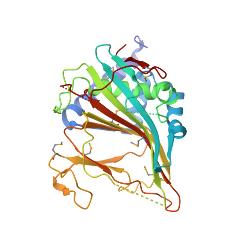
7EEH - PubMed Abstract:
Aziridine is a characteristically reactive molecule with increased bioactivity due to its strained ring structure. Here, we investigated the biosynthesis of 2-aminoisobutyric acid (AIB) in Penicillium, and successfully reconstituted the three-step biosynthesis from L-Val to AIB in vitro. This previously unknown aziridine formation pathway proceeded with the non-heme iron and α-ketoglutarate-dependent (Fe II /αKG) oxygenase TqaL, followed by aziridine ring opening by the haloalkanoic acid dehalogenase (HAD)-type hydrolase TqaF, and subsequent oxidative decarboxylation by the NovR/CloR-like non-heme iron oxygenase TqaM. Furthermore, the X-ray crystal structure of the C-N bond forming Fe II /αKG oxygenase TqaL was solved at 2.0 Å resolution. This work presents the first molecular basis for aziridine biogenesis, thereby expanding the catalytic repertoire of the Fe II /αKG oxygenases. We also report the unique aziridine ring opening by a HAD-type hydrolase and the remarkable oxidative decarboxylation by a non-heme iron oxygenase to produce AIB.
- Graduate School of Pharmaceutical Sciences, The University of Tokyo, 7-3-1 Hongo, Bunkyo-ku, Tokyo, 113-0033, Japan.
Organizational Affiliation: