7ECS | pdb_00007ecs

Crystal Structure of Aspergillus terreus Glutamate Dehydrogenase (AtGDH) Complexed With Malonate and NADPH


Experimental Data Snapshot

  • Method: X-RAY DIFFRACTION
  • Resolution: 1.74 Å
  • R-Value Free: 
    0.163 (Depositor), 0.174 (DCC) 
  • R-Value Work: 
    0.139 (Depositor), 0.152 (DCC) 
  • R-Value Observed: 
    0.140 (Depositor) 

Starting Model: experimental
View more details

wwPDB Validation 3D Report Full Report

Validation slider image for 7ECS

Ligand Structure Quality Assessment 


This is version 1.2 of the entry. See complete history

Literature

Molecular insights into the inhibition of glutamate dehydrogenase by the dicarboxylic acid metabolites.

Godsora, B.K.J.Prakash, P.Punekar, N.S.Bhaumik, P.

(2022) Proteins 90: 810-823

  • DOI: https://doi.org/10.1002/prot.26276
  • Primary Citation Related Structures: 
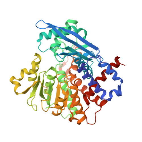
    7ECR, 7ECS, 7ECT

  • PubMed Abstract: 

    Glutamate dehydrogenase (GDH) is a salient metabolic enzyme which catalyzes the NAD + - or NADP + -dependent reversible conversion of α-ketoglutarate (AKG) to l-glutamate; and thereby connects the carbon and nitrogen metabolism cycles in all living organisms. The function of GDH is extensively regulated by both metabolites (citrate, succinate, etc.) and non-metabolites (ATP, NADH, etc.) but sufficient molecular evidences are lacking to rationalize the inhibitory effects by the metabolites. We have expressed and purified NADP + -dependent Aspergillus terreus GDH (AtGDH) in recombinant form. Succinate, malonate, maleate, fumarate, and tartrate independently inhibit the activity of AtGDH to different extents. The crystal structures of AtGDH complexed with the dicarboxylic acid metabolites and the coenzyme NADPH have been determined. Although AtGDH structures are not complexed with substrate; surprisingly, they acquire super closed conformation like previously reported for substrate and coenzyme bound catalytically competent Aspergillus niger GDH (AnGDH). These dicarboxylic acid metabolites partially occupy the same binding pocket as substrate; but interact with varying polar interactions and the coenzyme NADPH binds to the Domain-II of AtGDH. The low inhibition potential of tartrate as compared to other dicarboxylic acid metabolites is due to its weaker interactions of carboxylate groups with AtGDH. Our results suggest that the length of carbon skeleton and positioning of the carboxylate groups of inhibitors between two conserved lysine residues at the GDH active site might be the determinants of their inhibitory potency. Molecular details on the dicarboxylic acid metabolites bound AtGDH active site architecture presented here would be applicable to GDHs in general.


  • Organizational Affiliation
    • Department of Biosciences and Bioengineering, Indian Institute of Technology Bombay, Mumbai, Maharashtra, India.

Macromolecule Content 

  • Total Structure Weight: 152.8 kDa 
  • Atom Count: 12,647 
  • Modeled Residue Count: 1,380 
  • Deposited Residue Count: 1,380 
  • Unique protein chains: 1

Macromolecules

Find similar proteins by:|  3D Structure
Entity ID: 1
MoleculeChains  Sequence LengthOrganismDetailsImage
Glutamate dehydrogenase
A, B, C
460Aspergillus terreusMutation(s): 0 
Gene Names: gdhAATETN484_0007063400
UniProt
Find proteins for T2D1F5 (Aspergillus terreus)
Explore T2D1F5 
Go to UniProtKB:  T2D1F5
Entity Groups
Sequence Clusters30% Identity50% Identity70% Identity90% Identity95% Identity100% Identity
UniProt GroupT2D1F5
Sequence Annotations
Expand
Reference Sequence

Small Molecules

Ligands 3 Unique
IDChains Name / Formula / InChI Key2D Diagram3D Interactions
NDP
(Subject of Investigation/LOI)

Query on NDP


Download:Ideal Coordinates CCD File
JA [auth C],
M [auth A],
Y [auth B]
NADPH DIHYDRO-NICOTINAMIDE-ADENINE-DINUCLEOTIDE PHOSPHATE
C21 H30 N7 O17 P3
ACFIXJIJDZMPPO-NNYOXOHSSA-N
MLI
(Subject of Investigation/LOI)

Query on MLI


Download:Ideal Coordinates CCD File
HA [auth C]
IA [auth C]
K [auth A]
L [auth A]
W [auth B]
HA [auth C],
IA [auth C],
K [auth A],
L [auth A],
W [auth B],
X [auth B]
MALONATE ION
C3 H2 O4
OFOBLEOULBTSOW-UHFFFAOYSA-L
GOL

Query on GOL


Download:Ideal Coordinates CCD File
AA [auth C]
BA [auth C]
CA [auth C]
D [auth A]
DA [auth C]
AA [auth C],
BA [auth C],
CA [auth C],
D [auth A],
DA [auth C],
E [auth A],
EA [auth C],
F [auth A],
FA [auth C],
G [auth A],
GA [auth C],
H [auth A],
I [auth A],
J [auth A],
N [auth B],
O [auth B],
P [auth B],
Q [auth B],
R [auth B],
S [auth B],
T [auth B],
U [auth B],
V [auth B],
Z [auth C]
GLYCEROL
C3 H8 O3
PEDCQBHIVMGVHV-UHFFFAOYSA-N

Experimental Data & Validation

Experimental Data

  • Method: X-RAY DIFFRACTION
  • Resolution: 1.74 Å
  • R-Value Free:  0.163 (Depositor), 0.174 (DCC) 
  • R-Value Work:  0.139 (Depositor), 0.152 (DCC) 
  • R-Value Observed: 0.140 (Depositor) 
Space Group: I 2 2 2
Unit Cell:
Length ( Å )Angle ( ˚ )
a = 120.15α = 90
b = 153.74β = 90
c = 259.88γ = 90
Software Package:
Software NamePurpose
REFMACrefinement
XDSdata reduction
XSCALEdata scaling
PHASERphasing

Structure Validation

View Full Validation Report


Ligand Structure Quality Assessment 


Entry History 

& Funding Information

Deposition Data


Funding OrganizationLocationGrant Number
Department of Biotechnology (DBT, India)India--

Revision History  (Full details and data files)

  • Version 1.0: 2021-12-08
    Type: Initial release
  • Version 1.1: 2022-02-23
    Changes: Database references
  • Version 1.2: 2023-11-29
    Changes: Data collection, Refinement description