7EA6 | pdb_00007ea6

Crystal structure of TCR-017 ectodomain


Experimental Data Snapshot

  • Method: X-RAY DIFFRACTION
  • Resolution: 2.18 Å
  • R-Value Free: 
    0.276 (Depositor), 0.276 (DCC) 
  • R-Value Work: 
    0.222 (Depositor), 0.222 (DCC) 
  • R-Value Observed: 
    0.224 (Depositor) 

Starting Model: experimental
View more details

wwPDB Validation 3D Report Full Report

Validation slider image for 7EA6

This is version 1.3 of the entry. See complete history

Literature

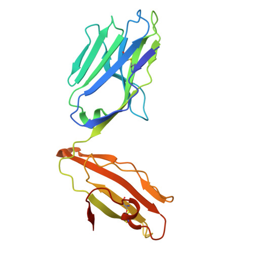
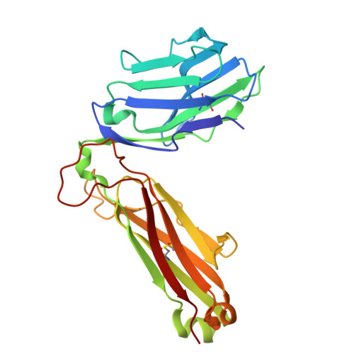
Identification of conserved SARS-CoV-2 spike epitopes that expand public cTfh clonotypes in mild COVID-19 patients.

Lu, X.Hosono, Y.Nagae, M.Ishizuka, S.Ishikawa, E.Motooka, D.Ozaki, Y.Sax, N.Maeda, Y.Kato, Y.Morita, T.Shinnakasu, R.Inoue, T.Onodera, T.Matsumura, T.Shinkai, M.Sato, T.Nakamura, S.Mori, S.Kanda, T.Nakayama, E.E.Shioda, T.Kurosaki, T.Takeda, K.Kumanogoh, A.Arase, H.Nakagami, H.Yamashita, K.Takahashi, Y.Yamasaki, S.

(2021) J Exp Medicine 218

  • DOI: https://doi.org/10.1084/jem.20211327
  • Primary Citation Related Structures: 
    7EA6

  • PubMed Abstract: 

    Adaptive immunity is a fundamental component in controlling COVID-19. In this process, follicular helper T (Tfh) cells are a subset of CD4+ T cells that mediate the production of protective antibodies; however, the SARS-CoV-2 epitopes activating Tfh cells are not well characterized. Here, we identified and crystallized TCRs of public circulating Tfh (cTfh) clonotypes that are expanded in patients who have recovered from mild symptoms. These public clonotypes recognized the SARS-CoV-2 spike (S) epitopes conserved across emerging variants. The epitope of the most prevalent cTfh clonotype, S864-882, was presented by multiple HLAs and activated T cells in most healthy donors, suggesting that this S region is a universal T cell epitope useful for booster antigen. SARS-CoV-2-specific public cTfh clonotypes also cross-reacted with specific commensal bacteria. In this study, we identified conserved SARS-CoV-2 S epitopes that activate public cTfh clonotypes associated with mild symptoms.


  • Organizational Affiliation
    • Laboratory of Molecular Immunology, Immunology Frontier Research Center, Osaka University, Suita, Japan.

Macromolecule Content 

  • Total Structure Weight: 99.3 kDa 
  • Atom Count: 7,161 
  • Modeled Residue Count: 865 
  • Deposited Residue Count: 880 
  • Unique protein chains: 2

Macromolecules

Find similar proteins by:|  3D Structure
Entity ID: 1
MoleculeChains  Sequence LengthOrganismDetailsImage
T cell receptor 017 alpha chainA [auth D],
C [auth A]
201Homo sapiensMutation(s): 0 
Find similar proteins by:|  3D Structure
Entity ID: 2
MoleculeChains  Sequence LengthOrganismDetailsImage
T cell receptor 017 beta chainB [auth E],
D [auth B]
239Homo sapiensMutation(s): 0 

Experimental Data & Validation

Experimental Data

  • Method: X-RAY DIFFRACTION
  • Resolution: 2.18 Å
  • R-Value Free:  0.276 (Depositor), 0.276 (DCC) 
  • R-Value Work:  0.222 (Depositor), 0.222 (DCC) 
  • R-Value Observed: 0.224 (Depositor) 
Space Group: C 2 2 21
Unit Cell:
Length ( Å )Angle ( ˚ )
a = 81.049α = 90
b = 81.175β = 90
c = 315.105γ = 90
Software Package:
Software NamePurpose
PHENIXdata reduction
PHENIXrefinement
Cootmodel building
SCALAdata scaling
MOLREPphasing

Structure Validation

View Full Validation Report


Entry History 

Deposition Data

Revision History  (Full details and data files)

  • Version 1.0: 2021-10-13
    Type: Initial release
  • Version 1.1: 2022-02-16
    Changes: Database references
  • Version 1.2: 2023-11-29
    Changes: Data collection, Refinement description
  • Version 1.3: 2024-11-06
    Changes: Structure summary