7E9I

Crystal structure of a class I PreQ1 riboswitch aptamer (wild-type) complexed with a cognate ligand-derived photoaffinity probe


Experimental Data Snapshot

  • Method: X-RAY DIFFRACTION
  • Resolution: 2.80 Å
  • R-Value Free: 0.235 
  • R-Value Work: 0.193 
  • R-Value Observed: 0.196 

wwPDB Validation   3D Report Full Report


Ligand Structure Quality Assessment 


This is version 1.1 of the entry. See complete history


Literature

A chemical probe based on the PreQ 1 metabolite enables transcriptome-wide mapping of binding sites.

Balaratnam, S.Rhodes, C.Bume, D.D.Connelly, C.Lai, C.C.Kelley, J.A.Yazdani, K.Homan, P.J.Incarnato, D.Numata, T.Schneekloth Jr., J.S.

(2021) Nat Commun 12: 5856-5856

  • DOI: https://doi.org/10.1038/s41467-021-25973-x
  • Primary Citation of Related Structures:  
    7E9E, 7E9I

  • PubMed Abstract: 

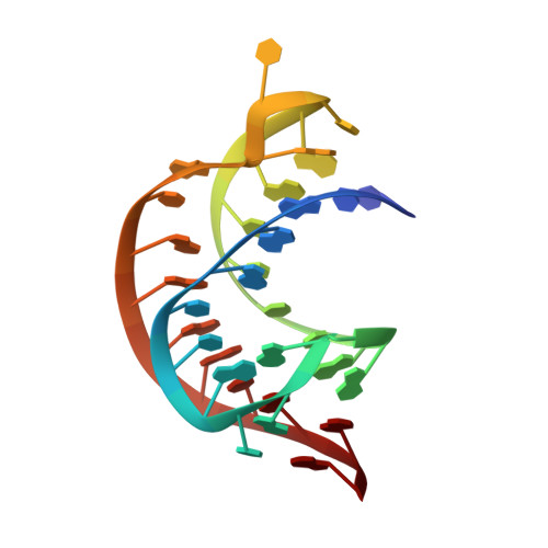
    The role of metabolite-responsive riboswitches in regulating gene expression in bacteria is well known and makes them useful systems for the study of RNA-small molecule interactions. Here, we study the PreQ 1 riboswitch system, assessing sixteen diverse PreQ 1 -derived probes for their ability to selectively modify the class-I PreQ 1 riboswitch aptamer covalently. For the most active probe (11), a diazirine-based photocrosslinking analog of PreQ 1 , X-ray crystallography and gel-based competition assays demonstrated the mode of binding of the ligand to the aptamer, and functional assays demonstrated that the probe retains activity against the full riboswitch. Transcriptome-wide mapping using Chem-CLIP revealed a highly selective interaction between the bacterial aptamer and the probe. In addition, a small number of RNA targets in endogenous human transcripts were found to bind specifically to 11, providing evidence for candidate PreQ 1 aptamers in human RNA. This work demonstrates a stark influence of linker chemistry and structure on the ability of molecules to crosslink RNA, reveals that the PreQ 1 aptamer/ligand pair are broadly useful for chemical biology applications, and provides insights into how PreQ 1 , which is similar in structure to guanine, interacts with human RNAs.


  • Organizational Affiliation

    Chemical Biology Laboratory, National Cancer Institute, Frederick, MD, 21702, USA.


Macromolecules
Find similar nucleic acids by:  (by identity cutoff)  |  3D Structure
Entity ID: 1
MoleculeChains LengthOrganismImage
33-mer RNA33Caldanaerobacter subterraneus subsp. tengcongensis
Sequence Annotations
Expand
  • Reference Sequence
Small Molecules
Ligands 2 Unique
IDChains Name / Formula / InChI Key2D Diagram3D Interactions
J0C
Query on J0C

Download Ideal Coordinates CCD File 
B [auth A]2-azanyl-5-[[2-(3-but-3-ynyl-1,2-diazirin-3-yl)ethylamino]methyl]-1,7-dihydropyrrolo[2,3-d]pyrimidin-4-one
C14 H17 N7 O
XAJHFEIBEFHTRZ-UHFFFAOYSA-N
SO4
Query on SO4

Download Ideal Coordinates CCD File 
C [auth A]SULFATE ION
O4 S
QAOWNCQODCNURD-UHFFFAOYSA-L
Experimental Data & Validation

Experimental Data

  • Method: X-RAY DIFFRACTION
  • Resolution: 2.80 Å
  • R-Value Free: 0.235 
  • R-Value Work: 0.193 
  • R-Value Observed: 0.196 
  • Space Group: P 63 2 2
Unit Cell:
Length ( Å )Angle ( ˚ )
a = 113.24α = 90
b = 113.24β = 90
c = 59.52γ = 120
Software Package:
Software NamePurpose
PHENIXrefinement
XDSdata reduction
XSCALEdata scaling
PHASERphasing

Structure Validation

View Full Validation Report


Ligand Structure Quality Assessment 


Entry History & Funding Information

Deposition Data


Funding OrganizationLocationGrant Number
Japan Society for the Promotion of Science (JSPS)Japan20K21281
Japan Society for the Promotion of Science (JSPS)Japan16KK0166
National Institutes of Health/National Cancer Institute (NIH/NCI)United States--

Revision History  (Full details and data files)

  • Version 1.0: 2021-11-17
    Type: Initial release
  • Version 1.1: 2023-11-29
    Changes: Data collection, Refinement description