7E7G | pdb_00007e7g

2-aminoethylphosphonate:pyruvate aminotransferase (AEPT) native


Experimental Data Snapshot

  • Method: X-RAY DIFFRACTION
  • Resolution: 2.35 Å
  • R-Value Free: 
    0.254 (Depositor), 0.227 (DCC) 
  • R-Value Work: 
    0.210 (Depositor), 0.206 (DCC) 

Starting Model: experimental
View more details

wwPDB Validation 3D Report Full Report

Validation slider image for 7E7G

This is version 1.1 of the entry. See complete history

Literature

Structural characterization of a 2-aminoethylphosphonate:pyruvate aminotransferase from Pseudomonas aeruginosa PAO1.

Jia, H.Chen, Y.Chen, Y.Liu, R.Zhang, Q.Bartlam, M.

(2021) Biochem Biophys Res Commun 552: 114-119

  • DOI: https://doi.org/10.1016/j.bbrc.2021.03.046
  • Primary Citation Related Structures: 
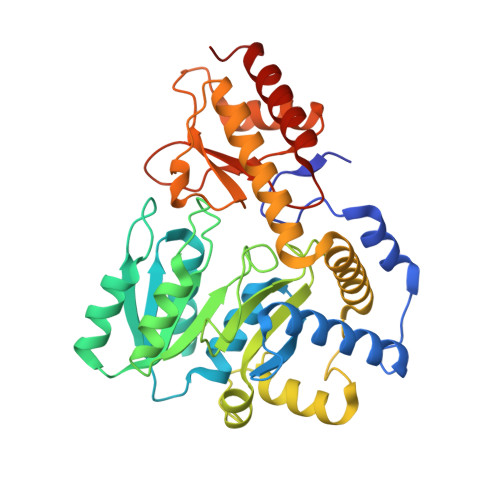
    7E7G

  • PubMed Abstract: 

    2-aminoethylphosphonate:pyruvate aminotransferase (AEPT) is a pyridoxal 5'-phosphate (PLP)-dependent enzyme that mediates the first step in the AEP degradation pathway. It catalyzes the transamination of 2-aminoethylphosphonate (AEP) with pyruvate to phosphonoacetaldehyde and l-alanine respectively. Although the enzyme is widely present in microorganisms, there are few reports on the structure and function of AEPT to date. Here we report the crystal structure of AEPT from Pseudomonas aeruginosa PAO1 (PaAEPT) to 2.35 Å resolution in the absence of the PLP cofactor. PaAEPT crystallizes in space group P2 1 2 1 2 with one monomer per asymmetric unit. Analytical ultracentrifugation analysis shows that PaAEPT forms a stable dimer in solution. Our work provides a valuable starting point for further functional and mechanistic studies of the AEP degradation pathway.


  • Organizational Affiliation
    • State Key Laboratory of Medicinal Chemical Biology, Tianjin Key Laboratory of Protein Science and College of Life Sciences, Nankai University, Tianjin, 300071, China.

Macromolecule Content 

  • Total Structure Weight: 40.47 kDa 
  • Atom Count: 2,770 
  • Modeled Residue Count: 362 
  • Deposited Residue Count: 371 
  • Unique protein chains: 1

Macromolecules

Find similar proteins by:|  3D Structure
Entity ID: 1
MoleculeChains  Sequence LengthOrganismDetailsImage
2-aminoethylphosphonate--pyruvate transaminase371Pseudomonas aeruginosa PAO1Mutation(s): 0 
Gene Names: phnWPA1310
EC: 2.6.1.37
UniProt
Find proteins for Q9I434 (Pseudomonas aeruginosa (strain ATCC 15692 / DSM 22644 / CIP 104116 / JCM 14847 / LMG 12228 / 1C / PRS 101 / PAO1))
Explore Q9I434 
Go to UniProtKB:  Q9I434
Entity Groups
Sequence Clusters30% Identity50% Identity70% Identity90% Identity95% Identity100% Identity
UniProt GroupQ9I434
Sequence Annotations
Expand
Reference Sequence

Experimental Data & Validation

Experimental Data

  • Method: X-RAY DIFFRACTION
  • Resolution: 2.35 Å
  • R-Value Free:  0.254 (Depositor), 0.227 (DCC) 
  • R-Value Work:  0.210 (Depositor), 0.206 (DCC) 
Space Group: P 21 21 2
Unit Cell:
Length ( Å )Angle ( ˚ )
a = 48.595α = 90
b = 69.207β = 90
c = 83.799γ = 90
Software Package:
Software NamePurpose
REFMACrefinement
HKL-3000data reduction
HKL-3000data scaling
PHASERphasing

Structure Validation

View Full Validation Report


Entry History 

& Funding Information

Deposition Data


Funding OrganizationLocationGrant Number
National Natural Science Foundation of China (NSFC)China31800627
National Natural Science Foundation of China (NSFC)China31870053

Revision History  (Full details and data files)

  • Version 1.0: 2021-03-31
    Type: Initial release
  • Version 1.1: 2023-11-29
    Changes: Data collection, Database references, Derived calculations, Refinement description