7E6H

glucose-6-phosphate dehydrogenase from Kluyveromyces lactis


Experimental Data Snapshot

  • Method: X-RAY DIFFRACTION
  • Resolution: 2.70 Å
  • R-Value Free: 0.237 
  • R-Value Work: 0.187 
  • R-Value Observed: 0.191 

wwPDB Validation   3D Report Full Report


This is version 1.1 of the entry. See complete history


Literature

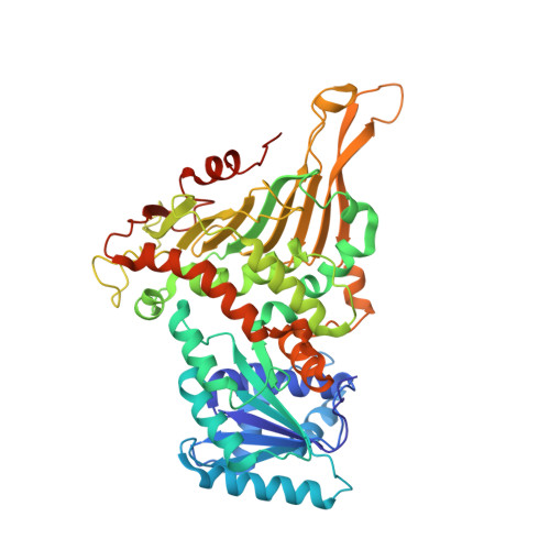
Structural basis for substrate recognition of glucose-6-phosphate dehydrogenase from Kluyveromyces lactis.

Vu, H.H.Jin, C.Chang, J.H.

(2021) Biochem Biophys Res Commun 553: 85-91

  • DOI: https://doi.org/10.1016/j.bbrc.2021.02.088
  • Primary Citation of Related Structures:  
    7E6H, 7E6I

  • PubMed Abstract: 

    Glucose-6-phosphate dehydrogenase is the first enzyme in the pentose phosphate pathway. The reaction catalyzed by the enzyme is considered to be the main source of reducing power for nicotinamide adenine dinucleotide phosphate (NADPH) and is a precursor of 5-carbon sugar used by cells. To uncover the structural features of the enzyme, we determined the crystal structures of glucose-6-phosphate dehydrogenase from Kluyveromyces lactis (KlG6PD) in both the apo form and a binary complex with its substrate glucose-6-phosphate. KlG6PD contains a Rossman-like domain for cofactor NADPH binding; it also presents a typical antiparallel β sheet at the C-terminal domain with relatively the same pattern as those of other homologous structures. Moreover, our structural and biochemical analyses revealed that Lys153 contributes significantly to substrate G6P recognition. This study may provide insights into the structural variation and catalytic features of the G6PD enzyme.


  • Organizational Affiliation

    Department of Biology Education, Kyungpook National University, Daegu, 41566, South Korea.


Macromolecules
Find similar proteins by:  (by identity cutoff)  |  3D Structure
Entity ID: 1
MoleculeChains Sequence LengthOrganismDetailsImage
Glucose-6-phosphate 1-dehydrogenase497Kluyveromyces lactis NRRL Y-1140Mutation(s): 0 
Gene Names: ZWFKLLA0D19855g
EC: 1.1.1.49
UniProt
Find proteins for P48828 (Kluyveromyces lactis (strain ATCC 8585 / CBS 2359 / DSM 70799 / NBRC 1267 / NRRL Y-1140 / WM37))
Explore P48828 
Go to UniProtKB:  P48828
Entity Groups  
Sequence Clusters30% Identity50% Identity70% Identity90% Identity95% Identity100% Identity
UniProt GroupP48828
Sequence Annotations
Expand
  • Reference Sequence
Small Molecules
Ligands 2 Unique
IDChains Name / Formula / InChI Key2D Diagram3D Interactions
7PE
Query on 7PE

Download Ideal Coordinates CCD File 
C [auth A]2-(2-(2-(2-(2-(2-ETHOXYETHOXY)ETHOXY)ETHOXY)ETHOXY)ETHOXY)ETHANOL
C14 H30 O7
UKXKPKBTMYNOFS-UHFFFAOYSA-N
TRS
Query on TRS

Download Ideal Coordinates CCD File 
B [auth A]2-AMINO-2-HYDROXYMETHYL-PROPANE-1,3-DIOL
C4 H12 N O3
LENZDBCJOHFCAS-UHFFFAOYSA-O
Experimental Data & Validation

Experimental Data

  • Method: X-RAY DIFFRACTION
  • Resolution: 2.70 Å
  • R-Value Free: 0.237 
  • R-Value Work: 0.187 
  • R-Value Observed: 0.191 
  • Space Group: P 65 2 2
Unit Cell:
Length ( Å )Angle ( ˚ )
a = 120.67α = 90
b = 120.67β = 90
c = 213.711γ = 120
Software Package:
Software NamePurpose
PHENIXrefinement
HKL-2000data reduction
HKL-2000data scaling
PHENIXphasing

Structure Validation

View Full Validation Report


Entry History & Funding Information

Deposition Data


Funding OrganizationLocationGrant Number
National Research Foundation (NRF, Korea)Korea, Republic OfNRF-2019R1A2C4069796

Revision History  (Full details and data files)

  • Version 1.0: 2021-04-14
    Type: Initial release
  • Version 1.1: 2023-11-29
    Changes: Data collection, Database references, Refinement description