


Structure-based inhibitor design for reshaping bacterial morphology
Choi, Y., Park, J.S., Kim, J., Min, K., Mahasenan, K., Kim, C., Yoon, H.J., Lim, S., Cheon, D.H., Lee, Y., Ryu, S., Mobashery, S., Kim, B.M., Lee, H.H.(2022) Commun Biol 5: 395
Experimental Data Snapshot
Starting Model: experimental
View more details
(2022) Commun Biol 5: 395
Entity ID: 1 | |||||
|---|---|---|---|---|---|
| Molecule | Chains | Sequence Length | Organism | Details | Image |
| Peptidase M23 | 255 | Campylobacter jejuni | Mutation(s): 1 Gene Names: A8118_01115 | 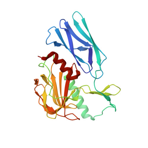 | |
| Ligands 2 Unique | |||||
|---|---|---|---|---|---|
| ID | Chains | Name / Formula / InChI Key | 2D Diagram | 3D Interactions | |
| HX6 (Subject of Investigation/LOI) Download:Ideal Coordinates CCD File | C [auth A] | N-oxidanyl-4-[(4-sulfamoylphenyl)methyl]benzamide C14 H14 N2 O4 S ZYVHYCNSGQRCBB-UHFFFAOYSA-N |  | ||
| ZN (Subject of Investigation/LOI) Download:Ideal Coordinates CCD File | B [auth A] | ZINC ION Zn PTFCDOFLOPIGGS-UHFFFAOYSA-N |  | ||
| Length ( Å ) | Angle ( ˚ ) |
|---|---|
| a = 115.649 | α = 90 |
| b = 115.649 | β = 90 |
| c = 56.614 | γ = 120 |
| Software Name | Purpose |
|---|---|
| XSCALE | data scaling |
| REFMAC | refinement |
| PDB_EXTRACT | data extraction |
| XDS | data reduction |
| MOLREP | phasing |


