


X-ray crystal structure of VapB12 antitoxin from mycobacterium tuberculosis in space group P41.
Pratap, S., Megta, A.K., Talwar, S., Sharma, C., Pandey, A.K., Krishnan, V.To be published.
Experimental Data Snapshot
wwPDB Validation 3D Report Full Report

Entity ID: 1 | |||||
|---|---|---|---|---|---|
| Molecule | Chains | Sequence Length | Organism | Details | Image |
| Antitoxin | 44 | Mycobacterium tuberculosis | Mutation(s): 0 Gene Names: vapB12, AYJ03_009075, DSI38_08650, E5M52_14165, E5M78_13900, ERS007663_00684, ERS007665_03495, ERS007703_02236, ERS007720_02286, ERS007722_03650... | 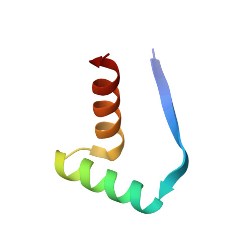 | |
UniProt | |||||
Entity Groups | |||||
| Sequence Clusters | 30% Identity50% Identity70% Identity90% Identity95% Identity100% Identity | ||||
| UniProt Group | P9WJ53 | ||||
Sequence AnnotationsExpand | |||||
Reference Sequence | |||||
| Ligands 1 Unique | |||||
|---|---|---|---|---|---|
| ID | Chains | Name / Formula / InChI Key | 2D Diagram | 3D Interactions | |
| ZN Download:Ideal Coordinates CCD File | E [auth B] | ZINC ION Zn PTFCDOFLOPIGGS-UHFFFAOYSA-N |  | ||
| Length ( Å ) | Angle ( ˚ ) |
|---|---|
| a = 34.66 | α = 90 |
| b = 34.66 | β = 90 |
| c = 162.227 | γ = 90 |
| Software Name | Purpose |
|---|---|
| REFMAC | refinement |
| XDS | data reduction |
| Aimless | data scaling |
| SHELXDE | phasing |


