


Crystal structure of histone lysine demethylase 4D (KDM4D) in complex with the inhibitor 5-hydroxy-2-methylpyrazolo[1,5-a]pyrido[3,2-e]pyrimidine-3-carbonitrile
Wang, T., Yang, L.To be published.
Experimental Data Snapshot
Starting Model: experimental
View more details
Entity ID: 1 | |||||
|---|---|---|---|---|---|
| Molecule | Chains | Sequence Length | Organism | Details | Image |
| Lysine-specific demethylase 4D | 330 | Homo sapiens | Mutation(s): 0 Gene Names: KDM4D, JHDM3D, JMJD2D EC: 1.14.11.66 | 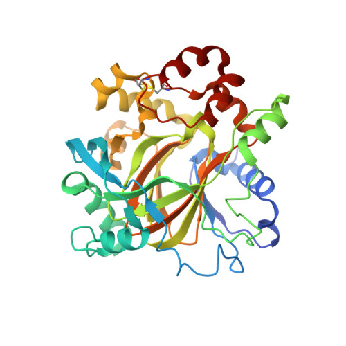 | |
UniProt & NIH Common Fund Data Resources | |||||
PHAROS: Q6B0I6 GTEx: ENSG00000186280 | |||||
Entity Groups | |||||
| Sequence Clusters | 30% Identity50% Identity70% Identity90% Identity95% Identity100% Identity | ||||
| UniProt Group | Q6B0I6 | ||||
Sequence AnnotationsExpand | |||||
Reference Sequence | |||||
| Ligands 2 Unique | |||||
|---|---|---|---|---|---|
| ID | Chains | Name / Formula / InChI Key | 2D Diagram | 3D Interactions | |
| HR0 (Subject of Investigation/LOI) Download:Ideal Coordinates CCD File | B [auth A] | 5-hydroxy-2-methylpyrazolo[1,5-a]pyrido[3,2-e]pyrimidine-3-carbonitrile C11 H7 N5 O FIRSAIIBSBCBTF-UHFFFAOYSA-N |  | ||
| FE Download:Ideal Coordinates CCD File | C [auth A] | FE (III) ION Fe VTLYFUHAOXGGBS-UHFFFAOYSA-N |  | ||
| Length ( Å ) | Angle ( ˚ ) |
|---|---|
| a = 71.635 | α = 90 |
| b = 71.635 | β = 90 |
| c = 151.486 | γ = 90 |
| Software Name | Purpose |
|---|---|
| PHENIX | refinement |
| PDB_EXTRACT | data extraction |


