Structural insights into how GlcNAc-1-phosphotransferase directs lysosomal protein transport.
Du, S., Wang, G., Zhang, Z., Ma, C., Gao, N., Xiao, J.(2022) J Biological Chem 298: 101702-101702
- PubMed: 35148990 Search on PubMedSearch on PubMed Central
- DOI: https://doi.org/10.1016/j.jbc.2022.101702
- Primary Citation Related Structures:
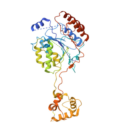
7DXI - PubMed Abstract:
GlcNAc-1-phosphotransferase catalyzes the initial step in the formation of the mannose-6-phosphate tag that labels ∼60 lysosomal proteins for transport. Mutations in GlcNAc-1-phosphotransferase are known to cause lysosomal storage disorders such as mucolipidoses. However, the molecular mechanism of GlcNAc-1-phosphotransferase activity remains unclear. Mammalian GlcNAc-1-phosphotransferases are α2β2γ2 hexamers in which the core catalytic α- and β-subunits are derived from the GNPTAB (N-acetylglucosamine-1-phosphate transferase subunits alpha and beta) gene. Here, we present the cryo-electron microscopy structure of the Drosophila melanogaster GNPTAB homolog, DmGNPTAB. We identified four conserved regions located far apart in the sequence that fold into the catalytic domain, which exhibits structural similarity to that of the UDP-glucose glycoprotein glucosyltransferase. Comparison with UDP-glucose glycoprotein glucosyltransferase also revealed a putative donor substrate-binding site, and the functional requirements of critical residues in human GNPTAB were validated using GNPTAB-knockout cells. Finally, we show that DmGNPTAB forms a homodimer that is evolutionarily conserved and that perturbing the dimer interface undermines the maturation and activity of human GNPTAB. These results provide important insights into GlcNAc-1-phosphotransferase function and related diseases.
- State Key Laboratory of Protein and Plant Gene Research, Peking University, Beijing, China; School of Life Sciences, Peking University, Beijing, China.
Organizational Affiliation: