7DRO

Structure of ATP-grasp ligase PsnB complexed with minimal precursor


Experimental Data Snapshot

  • Method: X-RAY DIFFRACTION
  • Resolution: 3.25 Å
  • R-Value Free: 0.263 
  • R-Value Work: 0.256 
  • R-Value Observed: 0.256 

wwPDB Validation   3D Report Full Report


This is version 1.3 of the entry. See complete history


Literature

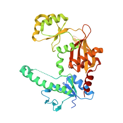
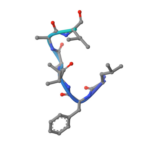
Molecular mechanism underlying substrate recognition of the peptide macrocyclase PsnB.

Song, I.Kim, Y.Yu, J.Go, S.Y.Lee, H.G.Song, W.J.Kim, S.

(2021) Nat Chem Biol 17: 1123-1131

  • DOI: https://doi.org/10.1038/s41589-021-00855-x
  • Primary Citation of Related Structures:  
    7DRM, 7DRN, 7DRO, 7DRP

  • PubMed Abstract: 

    Graspetides, also known as ω-ester-containing peptides (OEPs), are a family of ribosomally synthesized and post-translationally modified peptides (RiPPs) bearing side chain-to-side chain macrolactone or macrolactam linkages. Here, we present the molecular details of precursor peptide recognition by the macrocyclase enzyme PsnB in the biosynthesis of plesiocin, a group 2 graspetide. Biochemical analysis revealed that, in contrast to other RiPPs, the core region of the plesiocin precursor peptide noticeably enhanced the enzyme-precursor interaction via the conserved glutamate residues. We obtained four crystal structures of symmetric or asymmetric PsnB dimers, including those with a bound core peptide and a nucleotide, and suggest that the highly conserved Arg213 at the enzyme active site specifically recognizes a ring-forming acidic residue before phosphorylation. Collectively, this study provides insights into the mechanism underlying substrate recognition in graspetide biosynthesis and lays a foundation for engineering new variants.


  • Organizational Affiliation

    Department of Chemistry, Seoul National University, Seoul, South Korea.


Macromolecules
Find similar proteins by:  (by identity cutoff)  |  3D Structure
Entity ID: 1
MoleculeChains Sequence LengthOrganismDetailsImage
ATP-grasp domain-containing protein
A, B, C, D, E
A, B, C, D, E, F
334Plesiocystis pacifica SIR-1Mutation(s): 0 
Gene Names: PPSIR1_03893
UniProt
Find proteins for A6G4D7 (Plesiocystis pacifica SIR-1)
Explore A6G4D7 
Go to UniProtKB:  A6G4D7
Entity Groups  
Sequence Clusters30% Identity50% Identity70% Identity90% Identity95% Identity100% Identity
UniProt GroupA6G4D7
Sequence Annotations
Expand
  • Reference Sequence

Find similar proteins by:  Sequence   |   3D Structure  

Entity ID: 2
MoleculeChains Sequence LengthOrganismDetailsImage
PsnA214-38, Precursor peptide
G, H, I, J
25Plesiocystis pacifica SIR-1Mutation(s): 0 
UniProt
Find proteins for A6GH40 (Plesiocystis pacifica SIR-1)
Explore A6GH40 
Go to UniProtKB:  A6GH40
Entity Groups  
Sequence Clusters30% Identity50% Identity70% Identity90% Identity95% Identity100% Identity
UniProt GroupA6GH40
Sequence Annotations
Expand
  • Reference Sequence
Experimental Data & Validation

Experimental Data

  • Method: X-RAY DIFFRACTION
  • Resolution: 3.25 Å
  • R-Value Free: 0.263 
  • R-Value Work: 0.256 
  • R-Value Observed: 0.256 
  • Space Group: P 21 21 21
Unit Cell:
Length ( Å )Angle ( ˚ )
a = 86.894α = 90
b = 146.944β = 90
c = 167.834γ = 90
Software Package:
Software NamePurpose
PHENIXrefinement
HKL-2000data scaling
PDB_EXTRACTdata extraction
HKL-2000data reduction
PHENIXphasing

Structure Validation

View Full Validation Report


Entry History 

Deposition Data

Revision History  (Full details and data files)

  • Version 1.0: 2021-09-08
    Type: Initial release
  • Version 1.1: 2021-09-22
    Changes: Database references
  • Version 1.2: 2022-02-16
    Changes: Database references
  • Version 1.3: 2023-11-29
    Changes: Data collection, Refinement description