7DPH | pdb_00007dph

H-Ras Q61H in complex with GppNHp (state 1) after structural transition by humidity control


Experimental Data Snapshot

  • Method: X-RAY DIFFRACTION
  • Resolution: 1.54 Å
  • R-Value Free: 
    0.202 (Depositor), 0.213 (DCC) 
  • R-Value Work: 
    0.192 (Depositor), 0.202 (DCC) 

Starting Model: experimental
View more details

wwPDB Validation 3D Report Full Report

Validation slider image for 7DPH

Ligand Structure Quality Assessment 


This is version 1.1 of the entry. See complete history

Literature

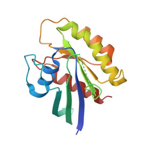
Oncogenic mutations Q61L and Q61H confer active form-like structural features to the inactive state (state 1) conformation of H-Ras protein.

Matsumoto, S.Taniguchi-Tamura, H.Araki, M.Kawamura, T.Miyamoto, R.Tsuda, C.Shima, F.Kumasaka, T.Okuno, Y.Kataoka, T.

(2021) Biochem Biophys Res Commun 565: 85-90

  • DOI: https://doi.org/10.1016/j.bbrc.2021.05.084
  • Primary Citation Related Structures: 
    7DPH, 7DPJ

  • PubMed Abstract: 

    GTP-bound forms of Ras proteins (Ras•GTP) assume two interconverting conformations, "inactive" state 1 and "active" state 2. Our previous study on the crystal structure of the state 1 conformation of H-Ras in complex with guanosine 5'-(β, γ-imido)triphosphate (GppNHp) indicated that state 1 is stabilized by intramolecular hydrogen-bonding interactions formed by Gln61. Since Ras are constitutively activated by substitution mutations of Gln61, here we determine crystal structures of the state 1 conformation of H-Ras•GppNHp carrying representative mutations Q61L and Q61H to observe the effect of the mutations. The results show that these mutations alter the mode of hydrogen-bonding interactions of the residue 61 with Switch II residues and induce conformational destabilization of the neighboring regions. In particular, Q61L mutation results in acquirement of state 2-like structural features. Moreover, the mutations are likely to impair an intramolecular structural communication between Switch I and Switch II. Molecular dynamics simulations starting from these structures support the above observations. These findings may give a new insight into the molecular mechanism underlying the aberrant activation of the Gln61 mutants.


  • Organizational Affiliation
    • Department of Biomedical Data Intelligence, Graduate School of Medicine, Kyoto University, 53 Shogoin-Kawaharacho, Sakyo-ku, Kyoto, 606-8507, Japan.

Macromolecule Content 

  • Total Structure Weight: 19.89 kDa 
  • Atom Count: 1,486 
  • Modeled Residue Count: 158 
  • Deposited Residue Count: 171 
  • Unique protein chains: 1

Macromolecules

Find similar proteins by:|  3D Structure
Entity ID: 1
MoleculeChains  Sequence LengthOrganismDetailsImage
GTPase HRas171Homo sapiensMutation(s): 1 
Gene Names: HRASHRAS1
EC: 3.6.5.2
UniProt & NIH Common Fund Data Resources
Find proteins for P01112 (Homo sapiens)
Explore P01112 
Go to UniProtKB:  P01112
PHAROS:  P01112
GTEx:  ENSG00000174775 
Entity Groups
Sequence Clusters30% Identity50% Identity70% Identity90% Identity95% Identity100% Identity
UniProt GroupP01112
Sequence Annotations
Expand
Reference Sequence

Experimental Data & Validation

Experimental Data

  • Method: X-RAY DIFFRACTION
  • Resolution: 1.54 Å
  • R-Value Free:  0.202 (Depositor), 0.213 (DCC) 
  • R-Value Work:  0.192 (Depositor), 0.202 (DCC) 
Space Group: H 3 2
Unit Cell:
Length ( Å )Angle ( ˚ )
a = 91.769α = 90
b = 91.769β = 90
c = 121.268γ = 120
Software Package:
Software NamePurpose
REFMACrefinement
HKL-2000data reduction
HKL-2000data scaling
MOLREPphasing

Structure Validation

View Full Validation Report


Ligand Structure Quality Assessment 


Entry History 

& Funding Information

Deposition Data


Funding OrganizationLocationGrant Number
Japan Society for the Promotion of Science (JSPS)JapanJP17K15106

Revision History  (Full details and data files)

  • Version 1.0: 2021-07-28
    Type: Initial release
  • Version 1.1: 2023-11-29
    Changes: Data collection, Database references, Derived calculations, Refinement description