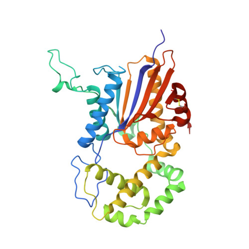
7DOQ | pdb_00007doq

Lp major histidine acid phosphatase mutant D281A/5'-AMP


Experimental Data Snapshot

  • Method: X-RAY DIFFRACTION
  • Resolution: 2.20 Å
  • R-Value Free: 
    0.229 (Depositor), 0.233 (DCC) 
  • R-Value Work: 
    0.192 (Depositor), 0.196 (DCC) 
  • R-Value Observed: 
    0.194 (Depositor) 

Starting Model: experimental
View more details

wwPDB Validation 3D Report Full Report

Validation slider image for 7DOQ

This is version 1.3 of the entry. See complete history. 

Literature

Structural insights into a new substrate binding mode of a histidine acid phosphatase from Legionella pneumophila.

Guo, Y., Zhou, D., Zhang, H., Zhang, N.N., Qi, X., Chen, X., Chen, Q., Li, J., Ge, H., Teng, Y.B.

(2021) Biochem Biophys Res Commun 540: 90-94

  • DOI: https://doi.org/10.1016/j.bbrc.2020.12.070
  • Primary Citation Related Structures: 
    7D2F, 7DOQ

  • PubMed Abstract: 

    MapA is a histidine acid phosphatase (HAP) from Legionella pneumophila that catalyzes the hydroxylation of a phosphoryl group from phosphomonoesters by an active-site histidine. Several structures of HAPs, including MapA, in complex with the inhibitor tartrate have been solved and the substrate binding tunnel identified; however, the substrate recognition mechanism remains unknown. To gain insight into the mechanism of substrate recognition, the crystal structures of apo-MapA and the MapA D281A mutant in complex with 5'-AMP were solved at 2.2 and 2.6 Å resolution, respectively. The structure of the MapA D281A /5'-AMP complex reveals that the 5'-AMP fits fully into the substrate binding tunnel, with the 2'-hydroxyl group of the ribose moiety stabilized by Glu201 and the adenine moiety sandwiched between His205 and Phe237. This is the second structure of a HAP/AMP complex solved with 5'-AMP binding in a unique manner in the active site. The structure presents a new substrate recognition mechanism of HAPs.


  • Organizational Affiliation: 
    • School of Life Sciences, Anhui Medical University, Hefei, Anhui, 230032, People's Republic of China. Electronic address: guoyu_joy@163.com.

Macromolecule Content 

  • Total Structure Weight: 151.49 kDa 
  • Atom Count: 10,841 
  • Modeled Residue Count: 1,328 
  • Deposited Residue Count: 1,340 
  • Unique protein chains: 1

Macromolecules

Find similar proteins by:|  3D Structure
Entity ID: 1
MoleculeChains  Sequence LengthOrganismDetailsImage
Acid phosphatase
A, B, C, D
335Legionella pneumophilaMutation(s): 0 
Gene Names: C3927_05010, DI026_02700
UniProt
Find proteins for Q9APF7 (Legionella pneumophila)
Explore Q9APF7 
Go to UniProtKB:  Q9APF7
Entity Groups
Sequence Clusters30% Identity50% Identity70% Identity90% Identity95% Identity100% Identity
UniProt GroupQ9APF7
Sequence Annotations
Expand
Reference Sequence

Experimental Data & Validation

Experimental Data

  • Method: X-RAY DIFFRACTION
  • Resolution: 2.20 Å
  • R-Value Free:  0.229 (Depositor), 0.233 (DCC) 
  • R-Value Work:  0.192 (Depositor), 0.196 (DCC) 
  • R-Value Observed: 0.194 (Depositor) 
Space Group: P 1 21 1
Unit Cell:
Length ( Å )Angle ( ˚ )
a = 91.498α = 90
b = 56.479β = 110.01
c = 146.349γ = 90
Software Package:
Software NamePurpose
REFMACrefinement
HKL-2000data scaling
PDB_EXTRACTdata extraction
HKL-2000data reduction
PHENIXphasing

Structure Validation

View Full Validation Report


Entry History 

& Funding Information

Deposition Data


Funding OrganizationLocationGrant Number
National Natural Science Foundation of China (NSFC)China31700650

Revision History  (Full details and data files)

  • Version 1.0: 2021-12-22
    Type: Initial release
  • Version 1.1: 2022-07-06
    Changes: Database references
  • Version 1.2: 2023-11-29
    Changes: Data collection, Refinement description
  • Version 1.3: 2024-10-23
    Changes: Structure summary