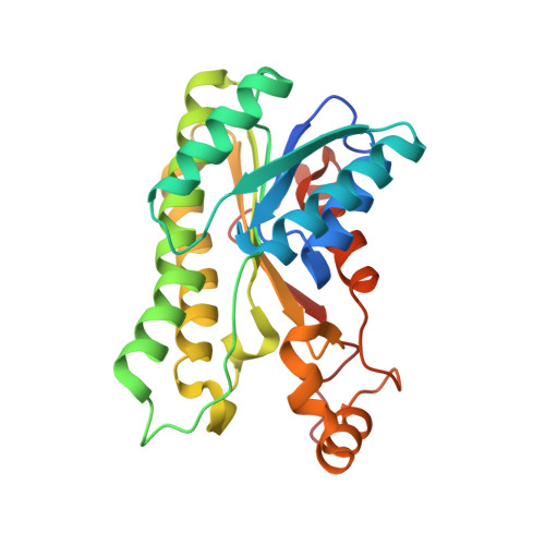
7DO7

Crystal structure of Azotobacter vinelandii L-rhamnose 1-dehydrogenase(NAD and L-rhamnose bound-form)


Experimental Data Snapshot

  • Method: X-RAY DIFFRACTION
  • Resolution: 1.57 Å
  • R-Value Free: 0.259 
  • R-Value Work: 0.223 
  • R-Value Observed: 0.224 

wwPDB Validation   3D Report Full Report


Ligand Structure Quality Assessment 


This is version 1.3 of the entry. See complete history


Literature

Crystal structure of l-rhamnose 1-dehydrogenase involved in the nonphosphorylative pathway of l-rhamnose metabolism in bacteria.

Yoshiwara, K.Watanabe, S.Watanabe, Y.

(2021) FEBS Lett 595: 637-646

  • DOI: https://doi.org/10.1002/1873-3468.14046
  • Primary Citation of Related Structures:  
    7B81, 7DO5, 7DO6, 7DO7

  • PubMed Abstract: 

    Several microorganisms can utilize l-rhamnose as a carbon and energy source through the nonphosphorylative metabolic pathway, in which l-rhamnose 1-dehydrogenase (RhaDH) catalyzes the NAD(P) + -dependent oxidization of l-rhamnose to l-rhamnono-1,4-lactone. We herein investigated the crystal structures of RhaDH from Azotobacter vinelandii in ligand-free, NAD + -bound, NADP + -bound, and l-rhamnose- and NAD + -bound forms at 1.9, 2.1, 2.4, and 1.6 Å resolution, respectively. The significant interactions with the 2'-phosphate group of NADP + , but not the 2'-hydroxyl group of NAD + , were consistent with a preference for NADP + over NAD + . The C5-OH and C6-methyl groups of l-rhamnose were recognized by specific residues of RhaDH through hydrogen bonds and hydrophobic contact, respectively, which contribute to the different substrate specificities from other aldose 1-dehydrogenases in the short-chain dehydrogenase/reductase superfamily.


  • Organizational Affiliation

    Faculty of Agriculture, Ehime University, Matsuyama, Japan.


Macromolecules
Find similar proteins by:  (by identity cutoff)  |  3D Structure
Entity ID: 1
MoleculeChains Sequence LengthOrganismDetailsImage
Short-chain dehydrogenase/reductase SDR
A, B
267Azotobacter vinelandii DJMutation(s): 0 
Gene Names: Avin_09160
UniProt
Find proteins for C1DMX5 (Azotobacter vinelandii (strain DJ / ATCC BAA-1303))
Explore C1DMX5 
Go to UniProtKB:  C1DMX5
Entity Groups  
Sequence Clusters30% Identity50% Identity70% Identity90% Identity95% Identity100% Identity
UniProt GroupC1DMX5
Sequence Annotations
Expand
  • Reference Sequence
Experimental Data & Validation

Experimental Data

  • Method: X-RAY DIFFRACTION
  • Resolution: 1.57 Å
  • R-Value Free: 0.259 
  • R-Value Work: 0.223 
  • R-Value Observed: 0.224 
  • Space Group: P 21 21 2
Unit Cell:
Length ( Å )Angle ( ˚ )
a = 68.47α = 90
b = 97.931β = 90
c = 70.302γ = 90
Software Package:
Software NamePurpose
PHENIXrefinement
XSCALEdata scaling
PDB_EXTRACTdata extraction
PHASERphasing

Structure Validation

View Full Validation Report


Ligand Structure Quality Assessment 


Entry History 

Deposition Data

Revision History  (Full details and data files)

  • Version 1.0: 2021-02-03
    Type: Initial release
  • Version 1.1: 2021-02-17
    Changes: Database references
  • Version 1.2: 2021-03-24
    Changes: Database references
  • Version 1.3: 2024-03-27
    Changes: Data collection, Database references