Functional and structural analysis of catabolite control protein C that responds to citrate.
Liu, W., Chen, J., Jin, L., Liu, Z.Y., Lu, M., Jiang, G., Yang, Q., Quan, C., Nam, K.H., Xu, Y.(2021) Sci Rep 11: 20285-20285
- PubMed: 34645869 Search on PubMedSearch on PubMed Central
- DOI: https://doi.org/10.1038/s41598-021-99552-x
- Primary Citation Related Structures:
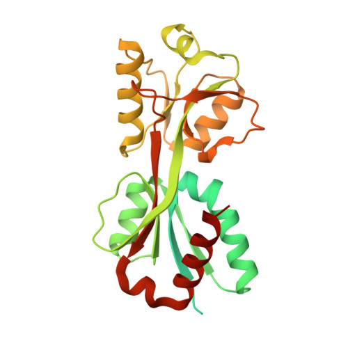
7DMW - PubMed Abstract:
Catabolite control protein C (CcpC) belongs to the LysR-type transcriptional regulator (LTTR) family, which regulates the transcription of genes encoding the tricarboxylic acid branch enzymes of the TCA cycle by responding to a pathway-specific metabolite, citrate. The biological function of CcpC has been characterized several times, but the structural basis for the molecular function of CcpC remains elusive. Here, we report the characterization of a full-length CcpC from Bacillus amyloliquefaciens (BaCcpC-FL) and a crystal structure of the C-terminal inducer-binding domain (IBD) complexed with citrate. BaCcpC required both dyad symmetric regions I and II to recognize the citB promoter, and the presence of citrate reduced citB promoter binding. The crystal structure of CcpC-IBD shows two subdomains, IBD-I and IBD-II, and a citrate molecule buried between them. Ile100, two arginines (Arg147 and Arg260), and three serines (Ser129, Ser189, and Ser191) exhibit strong hydrogen-bond interactions with citrate molecules. A structural comparison of BaCcpC-IBD with its homologues showed that they share the same tail-to-tail dimer alignment, but the dimeric interface and the rotation between these molecules exhibit significant differences. Taken together, our results provide a framework for understanding the mechanism underlying the functional divergence of the CcpC protein.
- Department of Bioengineering, College of Life Science, Dalian Minzu University, Dalian, 116600, Liaoning, China.
Organizational Affiliation: