7DMO | pdb_00007dmo

Crystal structures of two pericyclases catalyzing [4+2] cycloadditions


Experimental Data Snapshot

  • Method: X-RAY DIFFRACTION
  • Resolution: 2.00 Å
  • R-Value Free: 
    0.210 (Depositor), 0.193 (DCC) 
  • R-Value Work: 
    0.173 (Depositor) 
  • R-Value Observed: 
    0.173 (Depositor) 

Starting Model: experimental
View more details

wwPDB Validation 3D Report Full Report

Validation slider image for 7DMO

This is version 1.1 of the entry. See complete history

Literature

Crystal Structures of Fsa2 and Phm7 Catalyzing [4 + 2] Cycloaddition Reactions with Reverse Stereoselectivities in Equisetin and Phomasetin Biosynthesis.

Chi, C.Wang, Z.Liu, T.Zhang, Z.Zhou, H.Li, A.Jin, H.Jia, H.Yin, F.Yang, D.Ma, M.

(2021) ACS Omega 6: 12913-12922

  • DOI: https://doi.org/10.1021/acsomega.1c01593
  • Primary Citation Related Structures: 
    7DMN, 7DMO

  • PubMed Abstract: 

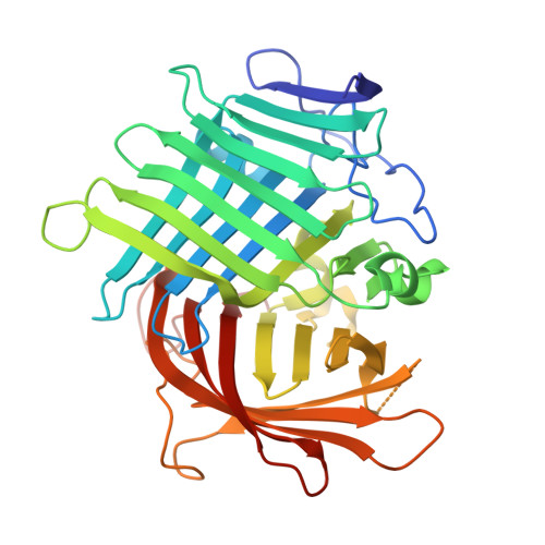
    Fsa2 and Phm7 are a unique pair of pericyclases catalyzing [4 + 2] cycloaddition reactions with reverse stereoselectivities in the biosynthesis of equisetin and phomasetin, both of which are potent HIV-1 integrase inhibitors. We here solve the crystal structures of Fsa2 and Phm7, both of which possess unusual "two-β barrel" folds. Different residues are evident between the active sites of Fsa2 and Phm7, and modeling experiments provide key structural information determining the reverse stereoselectivities. These results provide a better understanding of how natural pericyclases control the catalytic stereoselectivities and benefit the protein engineering in future.


  • Organizational Affiliation
    • State Key Laboratory of Natural and Biomimetic Drugs, School of Pharmaceutical Sciences, Peking University, 38 Xueyuan Road, Haidian District, Beijing 100191, China.

Macromolecule Content 

  • Total Structure Weight: 250.51 kDa 
  • Atom Count: 18,922 
  • Modeled Residue Count: 2,247 
  • Deposited Residue Count: 2,334 
  • Unique protein chains: 1

Macromolecules

Find similar proteins by:|  3D Structure
Entity ID: 1
MoleculeChains  Sequence LengthOrganismDetailsImage
Diels-Alderase
A, B, C, D, E
A, B, C, D, E, F
389Pyrenochaetopsis sp.Mutation(s): 0 
Gene Names: phm7
EC: 5.5.1
UniProt
Find proteins for A0A2Z5XAU0 (Pyrenochaetopsis sp)
Explore A0A2Z5XAU0 
Go to UniProtKB:  A0A2Z5XAU0
Entity Groups
Sequence Clusters30% Identity50% Identity70% Identity90% Identity95% Identity100% Identity
UniProt GroupA0A2Z5XAU0
Sequence Annotations
Expand
Reference Sequence

Experimental Data & Validation

Experimental Data

  • Method: X-RAY DIFFRACTION
  • Resolution: 2.00 Å
  • R-Value Free:  0.210 (Depositor), 0.193 (DCC) 
  • R-Value Work:  0.173 (Depositor) 
  • R-Value Observed: 0.173 (Depositor) 
Space Group: P 1 21 1
Unit Cell:
Length ( Å )Angle ( ˚ )
a = 91.196α = 90
b = 150.533β = 96.94
c = 100.047γ = 90
Software Package:
Software NamePurpose
PHENIXrefinement
PDB_EXTRACTdata extraction
CrysalisProdata reduction
CrysalisProdata scaling
PHENIXphasing

Structure Validation

View Full Validation Report


Entry History 

& Funding Information

Deposition Data


Funding OrganizationLocationGrant Number
Ministry of Science and Technology (MoST, China)China2019YFC0312502

Revision History  (Full details and data files)

  • Version 1.0: 2021-10-06
    Type: Initial release
  • Version 1.1: 2023-11-29
    Changes: Data collection, Refinement description