7DLS | pdb_00007dls

Cytochrome P450 (CYP105D18) complex with papaverine


Experimental Data Snapshot

  • Method: X-RAY DIFFRACTION
  • Resolution: 2.06 Å
  • R-Value Free: 
    0.218 (Depositor), 0.211 (DCC) 
  • R-Value Work: 
    0.172 (Depositor) 
  • R-Value Observed: 
    0.174 (Depositor) 

Starting Model: experimental
View more details

wwPDB Validation 3D Report Full Report

Validation slider image for 7DLS

Ligand Structure Quality Assessment 


This is version 1.2 of the entry. See complete history

Literature

Characterization of high-H 2 O 2 -tolerant bacterial cytochrome P450 CYP105D18: insights into papaverine N-oxidation.

Pardhe, B.D.Do, H.Jeong, C.S.Kim, K.H.Lee, J.H.Oh, T.J.

(2021) IUCrJ 8: 684-694

  • DOI: https://doi.org/10.1107/S2052252521005522
  • Primary Citation Related Structures: 
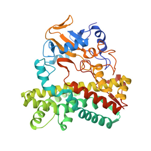
    7DI3, 7DLS

  • PubMed Abstract: 

    The bacterial CYP105 family is involved in secondary metabolite biosynthetic pathways and plays essential roles in the biotransformation of xenobiotics. This study investigates the newly identified H 2 O 2 -mediated CYP105D18 from Streptomyces laurentii as the first bacterial CYP for N-oxidation. The catalytic efficiency of CYP105D18 for papaverine N-oxidation was 1.43 s -1  µ M -1 . The heme oxidation rate ( k ) was low (<0.3 min -1 ) in the presence of 200 m M H 2 O 2 . This high H 2 O 2 tolerance capacity of CYP105D18 led to higher turnover prior to heme oxidation. Additionally, the high-resolution papaverine complexed structure and substrate-free structure of CYP105D18 were determined. Structural analysis and activity assay results revealed that CYP105D18 had a strong substrate preference for papaverine because of its bendable structure. These findings establish a basis for biotechnological applications of CYP105D18 in the pharmaceutical and medicinal industries.


  • Organizational Affiliation
    • Department of Life Science and Biochemical Engineering, Graduate School, SunMoon University, Asan 31460, Republic of Korea.

Macromolecule Content 

  • Total Structure Weight: 45.36 kDa 
  • Atom Count: 3,223 
  • Modeled Residue Count: 392 
  • Deposited Residue Count: 396 
  • Unique protein chains: 1

Macromolecules

Find similar proteins by:|  3D Structure
Entity ID: 1
MoleculeChains  Sequence LengthOrganismDetailsImage
Cytochrome P450 hydroxylase396Streptomyces laurentiiMutation(s): 0 
Gene Names: SLA_5925
UniProt
Find proteins for A0A160P685 (Streptomyces laurentii)
Explore A0A160P685 
Go to UniProtKB:  A0A160P685
Entity Groups
Sequence Clusters30% Identity50% Identity70% Identity90% Identity95% Identity100% Identity
UniProt GroupA0A160P685
Sequence Annotations
Expand
Reference Sequence

Small Molecules

Ligands 2 Unique
IDChains Name / Formula / InChI Key2D Diagram3D Interactions
HEM
(Subject of Investigation/LOI)

Query on HEM


Download:Ideal Coordinates CCD File
B [auth A]PROTOPORPHYRIN IX CONTAINING FE
C34 H32 Fe N4 O4
KABFMIBPWCXCRK-RGGAHWMASA-L
EV1
(Subject of Investigation/LOI)

Query on EV1


Download:Ideal Coordinates CCD File
C [auth A]1-(3,4-DIMETHOXYBENZYL)-6,7-DIMETHOXYISOQUINOLINE
C20 H21 N O4
XQYZDYMELSJDRZ-UHFFFAOYSA-N

Experimental Data & Validation

Experimental Data

  • Method: X-RAY DIFFRACTION
  • Resolution: 2.06 Å
  • R-Value Free:  0.218 (Depositor), 0.211 (DCC) 
  • R-Value Work:  0.172 (Depositor) 
  • R-Value Observed: 0.174 (Depositor) 
Space Group: C 1 2 1
Unit Cell:
Length ( Å )Angle ( ˚ )
a = 91.522α = 90
b = 52.172β = 110.68
c = 87.896γ = 90
Software Package:
Software NamePurpose
REFMACrefinement
PDB_EXTRACTdata extraction
HKL-2000data reduction
SCALAdata scaling
MOLREPphasing

Structure Validation

View Full Validation Report


Ligand Structure Quality Assessment 


Entry History 

Deposition Data

Revision History  (Full details and data files)

  • Version 1.0: 2021-07-14
    Type: Initial release
  • Version 1.1: 2021-07-28
    Changes: Database references
  • Version 1.2: 2023-11-29
    Changes: Data collection, Database references, Refinement description, Structure summary