Molecular basis for substrate recognition by the bacterial nucleoside transporter NupG.
Wang, C., Xiao, Q., Duan, H., Li, J., Zhang, J., Wang, Q., Guo, L., Hu, J., Sun, B., Deng, D.(2021) J Biol Chem 296: 100479-100479
- PubMed: 33640454 Search on PubMedSearch on PubMed Central
- DOI: https://doi.org/10.1016/j.jbc.2021.100479
- Primary Citation Related Structures:
7DL9, 7DLA - PubMed Abstract:
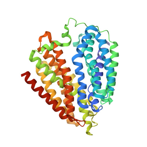
Nucleoside homeostasis, which is mediated by transporters and channels, is essential for all life on Earth. In Escherichia coli, NupG mediates the transport of nucleosides and was deemed to be the prototype of the nucleoside proton symporter (NHS) family and the major facilitator superfamily. To date, the substrate recognition and transport mechanisms of NHS transporters are still elusive. Here, we report two crystal structures of NupG (WT and D323A NupG) resolved at 3.0 Å. Both structures reveal an identical inward-open conformation. Together with molecular docking and molecular dynamics simulations and in vitro uridine-binding assays, we found that the uridine binding site, which locates in the central cavity between N and C domains of NupG, is constituted by R136, T140, F143, Q225, N228, Q261, E264, Y318, and F322. Moreover, we found that D323 is very important for substrate binding via in vitro uridine-binding assays using D323 mutations, although it does not have a direct contact with uridine. Our structural and biochemical data therefore provide an important framework for the mechanistic understanding of nucleoside transporters of the NHS family.
- Key Laboratory of Birth Defects and Related Disease of Women and Children of MOE, State Key Laboratory of Biotherapy, Department of Obstetrics, West China Second Hospital, Sichuan University, Chengdu, China.
Organizational Affiliation: