Protein and Organic-Molecular Crystallography With 300kV Electrons on a Direct Electron Detector.
Takaba, K., Maki-Yonekura, S., Inoue, S., Hasegawa, T., Yonekura, K.(2020) Front Mol Biosci 7: 612226-612226
- PubMed: 33469549 Search on PubMedSearch on PubMed Central
- DOI: https://doi.org/10.3389/fmolb.2020.612226
- Primary Citation Related Structures:
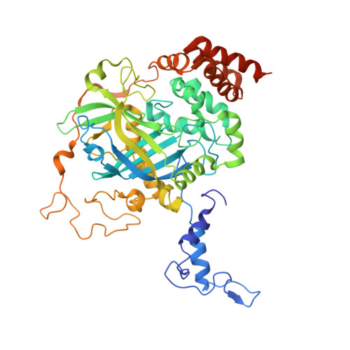
7DI8 - PubMed Abstract:
Electron 3D crystallography can reveal the atomic structure from undersized crystals of various samples owing to the strong scattering power of electrons. Here, a direct electron detector DE64 was tested for small and thin crystals of protein and an organic molecule using a JEOL CRYO ARM 300 electron microscope. The microscope is equipped with a cold-field emission gun operated at an accelerating voltage of 300 kV, quad condenser lenses for parallel illumination, an in-column energy filter, and a stable rotational goniometer stage. Rotational diffraction data were collected in an unsupervised manner from crystals of a heme-binding enzyme catalase and a representative organic semiconductor material Ph-BTBT-C10. The structures were determined by molecular replacement for catalase and by the direct method for Ph-BTBT-C10. The analyses demonstrate that the system works well for electron 3D crystallography of these molecules with less damaging, a smaller point spread, and less noise than using the conventional scintillator-coupled camera.
- Biostructural Mechanism Laboratory, RIKEN SPring-8 Center, Sayo, Japan.
Organizational Affiliation: