7DHY

Arsenic-bound p53 DNA-binding domain mutant G245S

  • Classification: TRANSCRIPTION
  • Organism(s): Homo sapiens
  • Expression System: Escherichia coli
  • Mutation(s): Yes 

  • Deposited: 2020-11-18 Released: 2021-03-03 
  • Deposition Author(s): Chen, S., Lu, M.
  • Funding Organization(s): Ministry of Science and Technology (MoST, China), National Natural Science Foundation of China (NSFC)

Experimental Data Snapshot

  • Method: X-RAY DIFFRACTION
  • Resolution: 2.15 Å
  • R-Value Free: 0.219 
  • R-Value Work: 0.187 
  • R-Value Observed: 0.189 

wwPDB Validation   3D Report Full Report


This is version 1.1 of the entry. See complete history


Literature

Arsenic Trioxide Rescues Structural p53 Mutations through a Cryptic Allosteric Site.

Chen, S.Wu, J.L.Liang, Y.Tang, Y.G.Song, H.X.Wu, L.L.Xing, Y.F.Yan, N.Li, Y.T.Wang, Z.Y.Xiao, S.J.Lu, X.Chen, S.J.Lu, M.

(2021) Cancer Cell 39: 225-239.e8

  • DOI: https://doi.org/10.1016/j.ccell.2020.11.013
  • Primary Citation of Related Structures:  
    7DHY, 7DHZ

  • PubMed Abstract: 

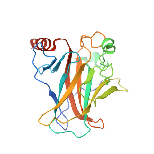
    TP53 is the most frequently mutated gene in cancer, yet these mutations remain therapeutically non-actionable. Major challenges in drugging p53 mutations include heterogeneous mechanisms of inactivation and the absence of broadly applicable allosteric sites. Here we report the identification of small molecules, including arsenic trioxide (ATO), an established agent in treating acute promyelocytic leukemia, as cysteine-reactive compounds that rescue structural p53 mutations. Crystal structures of arsenic-bound p53 mutants reveal a cryptic allosteric site involving three arsenic-coordinating cysteines within the DNA-binding domain, distal to the zinc-binding site. Arsenic binding stabilizes the DNA-binding loop-sheet-helix motif alongside the overall β-sandwich fold, endowing p53 mutants with thermostability and transcriptional activity. In cellular and mouse xenograft models, ATO reactivates mutant p53 for tumor suppression. Investigation of the 25 most frequent p53 mutations informs patient stratification for clinical exploration. Our results provide a mechanistic basis for repurposing ATO to target p53 mutations for widely applicable yet personalized cancer therapies.


  • Organizational Affiliation

    Shanghai Institute of Hematology, State Key Laboratory of Medical Genomics, National Research Center for Translational Medicine (Shanghai), Ruijin Hospital affiliated with Shanghai Jiao Tong University School of Medicine, Shanghai 200025, China; Ludwig Institute for Cancer Research, Nuffield Department of Medicine, University of Oxford, Oxford OX3 7DQ, UK.


Macromolecules
Find similar proteins by:  (by identity cutoff)  |  3D Structure
Entity ID: 1
MoleculeChains Sequence LengthOrganismDetailsImage
Cellular tumor antigen p53
A, B, C, D
200Homo sapiensMutation(s): 1 
Gene Names: TP53P53
UniProt & NIH Common Fund Data Resources
Find proteins for P04637 (Homo sapiens)
Explore P04637 
Go to UniProtKB:  P04637
PHAROS:  P04637
GTEx:  ENSG00000141510 
Entity Groups  
Sequence Clusters30% Identity50% Identity70% Identity90% Identity95% Identity100% Identity
UniProt GroupP04637
Sequence Annotations
Expand
  • Reference Sequence
Small Molecules
Ligands 4 Unique
IDChains Name / Formula / InChI Key2D Diagram3D Interactions
SO4
Query on SO4

Download Ideal Coordinates CCD File 
Q [auth D]SULFATE ION
O4 S
QAOWNCQODCNURD-UHFFFAOYSA-L
GOL
Query on GOL

Download Ideal Coordinates CCD File 
I [auth B]
J [auth B]
K [auth B]
N [auth C]
R [auth D]
I [auth B],
J [auth B],
K [auth B],
N [auth C],
R [auth D],
S [auth D],
T [auth D]
GLYCEROL
C3 H8 O3
PEDCQBHIVMGVHV-UHFFFAOYSA-N
ARS (Subject of Investigation/LOI)
Query on ARS

Download Ideal Coordinates CCD File 
E [auth A],
G [auth B],
L [auth C],
O [auth D]
ARSENIC
As
RBFQJDQYXXHULB-UHFFFAOYSA-N
ZN
Query on ZN

Download Ideal Coordinates CCD File 
F [auth A],
H [auth B],
M [auth C],
P [auth D]
ZINC ION
Zn
PTFCDOFLOPIGGS-UHFFFAOYSA-N
Experimental Data & Validation

Experimental Data

  • Method: X-RAY DIFFRACTION
  • Resolution: 2.15 Å
  • R-Value Free: 0.219 
  • R-Value Work: 0.187 
  • R-Value Observed: 0.189 
  • Space Group: P 1 21 1
Unit Cell:
Length ( Å )Angle ( ˚ )
a = 68.109α = 90
b = 68.243β = 90.03
c = 83.621γ = 90
Software Package:
Software NamePurpose
REFMACrefinement
PDB_EXTRACTdata extraction
XDSdata reduction
XDSdata scaling
PHASERphasing

Structure Validation

View Full Validation Report


Entry History & Funding Information

Deposition Data


Funding OrganizationLocationGrant Number
Ministry of Science and Technology (MoST, China)China2017YFA0506200
National Natural Science Foundation of China (NSFC)China81622002
National Natural Science Foundation of China (NSFC)China81861130368
National Natural Science Foundation of China (NSFC)China82073292

Revision History  (Full details and data files)

  • Version 1.0: 2021-03-03
    Type: Initial release
  • Version 1.1: 2023-11-29
    Changes: Data collection, Database references, Refinement description