


Structural insights into kinetoplastid coronin oligomerization domain and F-actin interaction
Parihar, P.S., Singh, A., Karade, S.S., Sahasrabuddhe, A.A., Pratap, J.V.(2021) Curr Res Struct Biol 3: 268-276
Experimental Data Snapshot
Starting Model: experimental
View more details
wwPDB Validation 3D Report Full Report

(2021) Curr Res Struct Biol 3: 268-276
Entity ID: 1 | |||||
|---|---|---|---|---|---|
| Molecule | Chains | Sequence Length | Organism | Details | Image |
| Coronin | 52 | Trypanosoma brucei | Mutation(s): 0 | 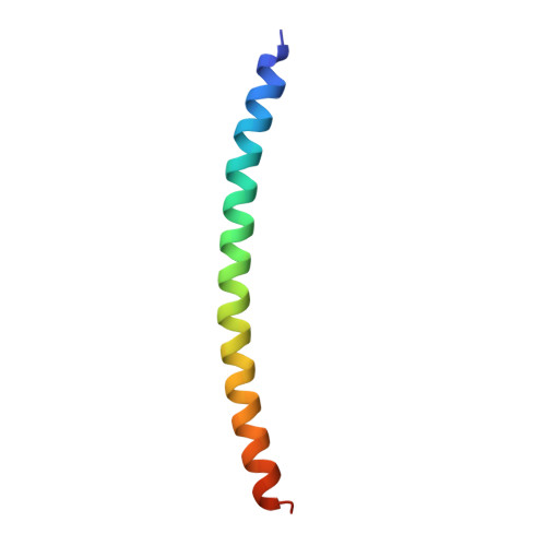 | |
UniProt | |||||
Entity Groups | |||||
| Sequence Clusters | 30% Identity50% Identity70% Identity90% Identity95% Identity100% Identity | ||||
| UniProt Group | Q57W63 | ||||
Sequence AnnotationsExpand | |||||
Reference Sequence | |||||
| Length ( Å ) | Angle ( ˚ ) |
|---|---|
| a = 93.376 | α = 90 |
| b = 93.376 | β = 90 |
| c = 82.959 | γ = 90 |
| Software Name | Purpose |
|---|---|
| PHENIX | refinement |
| DENZO | data reduction |
| HKL-2000 | data scaling |
| PHASER | phasing |


