7DFR

CRYSTAL STRUCTURES OF ESCHERICHIA COLI DIHYDROFOLATE REDUCTASE. THE NADP+ HOLOENZYME AND THE FOLATE(DOT)NADP+ TERNARY COMPLEX. SUBSTRATE BINDING AND A MODEL FOR THE TRANSITION STATE


Experimental Data Snapshot

  • Method: X-RAY DIFFRACTION
  • Resolution: 2.50 Å
  • R-Value Observed: 0.245 

wwPDB Validation   3D Report Full Report


This is version 1.3 of the entry. See complete history


Literature

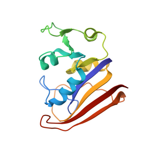
Crystal structures of Escherichia coli dihydrofolate reductase: the NADP+ holoenzyme and the folate.NADP+ ternary complex. Substrate binding and a model for the transition state.

Bystroff, C.Oatley, S.J.Kraut, J.

(1990) Biochemistry 29: 3263-3277

  • DOI: https://doi.org/10.1021/bi00465a018
  • Primary Citation of Related Structures:  
    6DFR, 7DFR

  • PubMed Abstract: 

    The crystal structure of dihydrofolate reductase (EC 1.5.1.3) from Escherichia coli has been solved as the binary complex with NADP+ (the holoenzyme) and as the ternary complex with NADP+ and folate. The Bragg law resolutions of the structures are 2.4 and 2.5 A, respectively. The new crystal forms are nonisomorphous with each other and with the methotrexate binary complex reported earlier [Bolin, J. T., Filman, D. J., Matthews, D. A., Hamlin, R. C., & Kraut, J. (1982) J. Biol. Chem. 257, 13650-13662]. In general, NADP+ and folate binding conform to predictions, but the nicotinamide moiety of NADP+ is disordered in the holoenzyme and ordered in the ternary complex. A mobile loop (residues 16-20) involved in binding the nicotinamide is also disordered in the holoenzyme. We report a detailed analysis of the binding interactions for both ligands, paying special attention to several apparently strained interactions that may favor the transition state for hydride transfer. Hypothetical models are presented for the binding of 7,8-dihydrofolate in the Michaelis complex and for the transition-state complex.


  • Organizational Affiliation

    Department of Chemistry, University of California, San Diego, La Jolla 92093.


Macromolecules
Find similar proteins by:  (by identity cutoff)  |  3D Structure
Entity ID: 1
MoleculeChains Sequence LengthOrganismDetailsImage
DIHYDROFOLATE REDUCTASE159Escherichia coliMutation(s): 0 
EC: 1.5.1.3
UniProt
Find proteins for P0ABQ4 (Escherichia coli (strain K12))
Explore P0ABQ4 
Go to UniProtKB:  P0ABQ4
Entity Groups  
Sequence Clusters30% Identity50% Identity70% Identity90% Identity95% Identity100% Identity
UniProt GroupP0ABQ4
Sequence Annotations
Expand
  • Reference Sequence
Small Molecules
Ligands 2 Unique
IDChains Name / Formula / InChI Key2D Diagram3D Interactions
NAP
Query on NAP

Download Ideal Coordinates CCD File 
C [auth A]NADP NICOTINAMIDE-ADENINE-DINUCLEOTIDE PHOSPHATE
C21 H28 N7 O17 P3
XJLXINKUBYWONI-NNYOXOHSSA-N
FOL
Query on FOL

Download Ideal Coordinates CCD File 
B [auth A]FOLIC ACID
C19 H19 N7 O6
OVBPIULPVIDEAO-LBPRGKRZSA-N
Experimental Data & Validation

Experimental Data

  • Method: X-RAY DIFFRACTION
  • Resolution: 2.50 Å
  • R-Value Observed: 0.245 
  • Space Group: P 32 2 1
Unit Cell:
Length ( Å )Angle ( ˚ )
a = 62.207α = 90
b = 62.207β = 90
c = 105.525γ = 120
Software Package:
Software NamePurpose
PROLSQrefinement

Structure Validation

View Full Validation Report


Entry History 

Deposition Data

Revision History  (Full details and data files)

  • Version 1.0: 1990-07-15
    Type: Initial release
  • Version 1.1: 2008-03-25
    Changes: Version format compliance
  • Version 1.2: 2011-07-13
    Changes: Version format compliance
  • Version 1.3: 2017-11-29
    Changes: Derived calculations, Other, Structure summary