


Crystal structure of M.tuberculosis imidazole glycerol phosphate dehydratase in complex with an inhibitor
Tiwari, S., Pal, R.K., Biswal, B.K.To be published.
Experimental Data Snapshot
Starting Model: experimental
View more details
Entity ID: 1 | |||||
|---|---|---|---|---|---|
| Molecule | Chains | Sequence Length | Organism | Details | Image |
| Imidazoleglycerol-phosphate dehydratase | 216 | Mycobacterium tuberculosis H37Rv | Mutation(s): 0 Gene Names: hisB, Rv1601, MTCY336.03c EC: 4.2.1.19 | 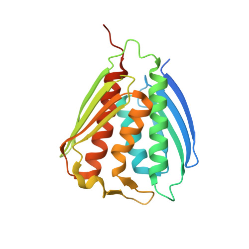 | |
UniProt | |||||
Entity Groups | |||||
| Sequence Clusters | 30% Identity50% Identity70% Identity90% Identity95% Identity100% Identity | ||||
| UniProt Group | P9WML9 | ||||
Sequence AnnotationsExpand | |||||
Reference Sequence | |||||
| Ligands 4 Unique | |||||
|---|---|---|---|---|---|
| ID | Chains | Name / Formula / InChI Key | 2D Diagram | 3D Interactions | |
| H3L (Subject of Investigation/LOI) Download:Ideal Coordinates CCD File | D [auth A] | (1S)-1-(2-methyl-1,2,4-triazol-3-yl)ethanamine C5 H10 N4 OCXIKYJPCILJLE-BYPYZUCNSA-N |  | ||
| GOL Download:Ideal Coordinates CCD File | E [auth A] | GLYCEROL C3 H8 O3 PEDCQBHIVMGVHV-UHFFFAOYSA-N |  | ||
| MN Download:Ideal Coordinates CCD File | B [auth A], C [auth A] | MANGANESE (II) ION Mn WAEMQWOKJMHJLA-UHFFFAOYSA-N |  | ||
| CL Download:Ideal Coordinates CCD File | F [auth A], G [auth A] | CHLORIDE ION Cl VEXZGXHMUGYJMC-UHFFFAOYSA-M |  | ||
| Length ( Å ) | Angle ( ˚ ) |
|---|---|
| a = 111.829 | α = 90 |
| b = 111.829 | β = 90 |
| c = 111.829 | γ = 90 |
| Software Name | Purpose |
|---|---|
| REFMAC | refinement |
| HKL-2000 | data reduction |
| HKL-2000 | data scaling |
| PHASER | phasing |


