7DC6 | pdb_00007dc6

Giant panda MHC class I complexes


Experimental Data Snapshot

  • Method: X-RAY DIFFRACTION
  • Resolution: 2.68 Å
  • R-Value Free: 
    0.291 (Depositor), 0.291 (DCC) 
  • R-Value Work: 
    0.266 (Depositor), 0.266 (DCC) 
  • R-Value Observed: 
    0.268 (Depositor) 

Starting Model: experimental
View more details

wwPDB Validation 3D Report Full Report

Validation slider image for 7DC6

This is version 1.2 of the entry. See complete history

Literature

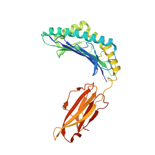
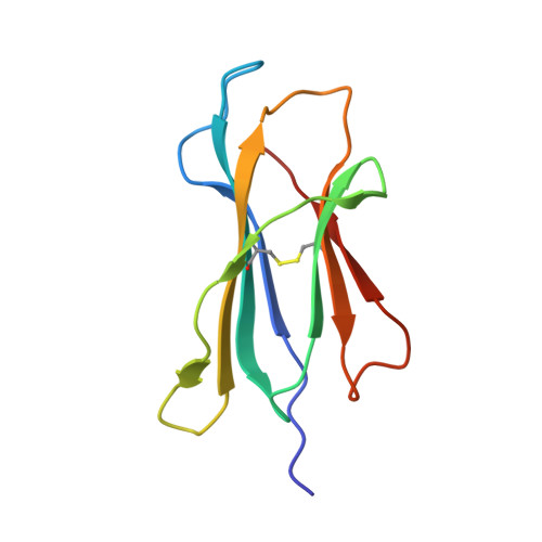
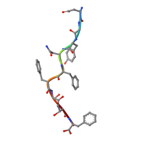
Crystal structure of the giant panda MHC class I complex: First insights into the viral peptide presentation profile in the bear family.

Yuan, H.Ma, L.Zhang, L.Li, X.Xia, C.

(2020) Protein Sci 29: 2468-2481

  • DOI: https://doi.org/10.1002/pro.3980
  • Primary Citation Related Structures: 
    7DC6

  • PubMed Abstract: 

    The viral cytotoxic T lymphocyte (CTL) epitope peptides presented by classical MHC-I molecules require the assembly of a peptide-MHC-I-β2m (pMHC-I) trimolecular complex for T cell receptor (TCR) recognition, which is the critical activation link for triggering antiviral T cell immunity. Research on T cell immunology in the Ursidae family, especially structural immunology, is still lacking. In this study, the structure of the key trimolecular complex pMHC-I, which binds a peptide from canine distemper virus, was solved for the first time using giant panda as a representative species of Ursidae. The structural characteristics of the giant panda pMHC-I complex (pAime-128), including the unique pockets in the peptide-binding groove (PBG), were analyzed in detail. Comparing the pAime-128 to others in the bear family and extending the comparison to other mammals revealed distinct features. The interaction between MHC-I and β2m, the features of pAime-128 involved in TCR docking and cluster of differentiation 8 (CD8) binding, the anchor sites in the PBG, and the CTL epitopes of potential viruses that infect pandas were clarified. Unique features of pMHC-I viral antigen presentation in the panda were revealed by solving the three-dimensional (3D) structure of pAime-128. The distinct characteristics of pAime-128 indicate an unusual event that emerged during the evolution of the MHC system in the bear family. These results provide a new platform for research on panda CTL immunity and the design of vaccines for application in the bear family.


  • Organizational Affiliation
    • Department of Microbiology and Immunology, College of Veterinary Medicine, China Agricultural University, Beijing, China.

Macromolecule Content 

  • Total Structure Weight: 88.9 kDa 
  • Atom Count: 6,298 
  • Modeled Residue Count: 765 
  • Deposited Residue Count: 766 
  • Unique protein chains: 3

Macromolecules

Find similar proteins by:|  3D Structure
Entity ID: 1
MoleculeChains  Sequence LengthOrganismDetailsImage
MHC class I antigen
A, C
275Ailuropoda melanoleucaMutation(s): 0 
Gene Names: Aime-128
UniProt
Find proteins for B2KT53 (Ailuropoda melanoleuca)
Explore B2KT53 
Go to UniProtKB:  B2KT53
Entity Groups
Sequence Clusters30% Identity50% Identity70% Identity90% Identity95% Identity100% Identity
UniProt GroupB2KT53
Sequence Annotations
Expand
Reference Sequence
Find similar proteins by:|  3D Structure
Entity ID: 2
MoleculeChains  Sequence LengthOrganismDetailsImage
Beta-2-microglobulin
B, D
99Ailuropoda melanoleucaMutation(s): 0 
Gene Names: B2MPANDA_000996
UniProt
Find proteins for A0A7N5K2L4 (Ailuropoda melanoleuca)
Explore A0A7N5K2L4 
Go to UniProtKB:  A0A7N5K2L4
Entity Groups
Sequence Clusters30% Identity50% Identity70% Identity90% Identity95% Identity100% Identity
UniProt GroupA0A7N5K2L4
Sequence Annotations
Expand
Reference Sequence
Find similar proteins by:  Sequence   |   3D Structure  
Entity ID: 3
MoleculeChains  Sequence LengthOrganismDetailsImage
CCV-NGY9 peptide from Spike protein
E, F
9Canine coronavirusMutation(s): 0 
UniProt
Find proteins for P10033 (Feline coronavirus (strain FIPV WSU-79/1146))
Explore P10033 
Go to UniProtKB:  P10033
Entity Groups
UniProt GroupP10033
Sequence Annotations
Expand
Reference Sequence

Experimental Data & Validation

Experimental Data

  • Method: X-RAY DIFFRACTION
  • Resolution: 2.68 Å
  • R-Value Free:  0.291 (Depositor), 0.291 (DCC) 
  • R-Value Work:  0.266 (Depositor), 0.266 (DCC) 
  • R-Value Observed: 0.268 (Depositor) 
Space Group: P 43 21 2
Unit Cell:
Length ( Å )Angle ( ˚ )
a = 173.677α = 90
b = 173.677β = 90
c = 84.346γ = 90
Software Package:
Software NamePurpose
REFMACrefinement
PDB_EXTRACTdata extraction
HKL-2000data reduction
HKL-2000data scaling
REFMACphasing

Structure Validation

View Full Validation Report


Entry History 

& Funding Information

Deposition Data


Funding OrganizationLocationGrant Number
National Natural Science Foundation of China (NSFC)China31572493
National Natural Science Foundation of China (NSFC)China31972683

Revision History  (Full details and data files)

  • Version 1.0: 2021-06-16
    Type: Initial release
  • Version 1.1: 2023-11-29
    Changes: Data collection, Database references, Refinement description
  • Version 1.2: 2024-10-16
    Changes: Structure summary