Functional and structural characterization of Deinococcus radiodurans R1 MazEF toxin-antitoxin system, Dr0416-Dr0417.
Dhanasingh, I., Choi, E., Lee, J., Lee, S.H., Hwang, J.(2021) J Microbiol 59: 186-201
- PubMed: 33527318 Search on PubMedSearch on PubMed Central
- DOI: https://doi.org/10.1007/s12275-021-0523-z
- Primary Citation Related Structures:
7D28, 7D2M, 7D2N, 7D2P, 7D2Q - PubMed Abstract:
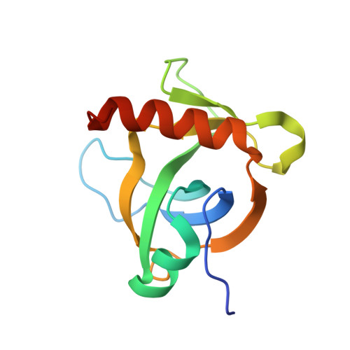
In prokaryotes, toxin-antitoxin (TA) systems are commonly found. They likely reflect the adaptation of pathogenic bacteria or extremophiles to various unfavorable environments by slowing their growth rate. Genomic analysis of the extremophile Deinococcus radiodurans R1 revealed the presence of eight type II TA systems, including the genes dr0417, dr0660, dr1530, dr0690, and dr1807. Expression of these toxin genes led to inhibition of Escherichia coli growth, whereas their antidote antitoxins were able to recover the growth defect. Remarkably, Dr0417 (DrMazF) showed endoribonuclease activity toward rRNAs as well as mRNAs, as determined by in vivo and in vitro RNA cleavage assays, and this activity was inhibited by Dr0416 (DrMazE). It was also found that the expression of dr0416-0417 module is directly regulated by the DrMazE-MazF complex. Furthermore, this TA module was induced under stress conditions and plays an important role in survival. To understand the regulatory mechanism at the molecular level, we determined the first high-resolution structures of DrMazF alone and of the DrMazE-MazF complex. In contrast with the hetero-hexameric state of typical MazE-MazF complexes found in other species, DrMazE-MazF crystal structure consists of a hetero-trimer, with the DNA-binding domain of DrMazE undergoing self-cleavage at the flexible linker loop. Our structure revealed that the unique residue R54 provides an additional positive charge to the substrate-binding pocket of DrMazF, its mutation significantly affects the endonuclease activity. Thus, our work reports the unique structural and biochemical features of the DrMazE-MazF system.
- Department of Cellular and Molecular Medicine, Chosun University School of Medicine, Gwangju, 61452, Republic of Korea.
Organizational Affiliation: