7D2K

Crystal structure of rat TRPV6 in complex with (4- phenylcyclohexyl)piperazine inhibitor Br-cis-22a


Experimental Data Snapshot

  • Method: X-RAY DIFFRACTION
  • Resolution: 3.70 Å
  • R-Value Free: 0.327 
  • R-Value Work: 0.316 
  • R-Value Observed: 0.317 

wwPDB Validation   3D Report Full Report


Ligand Structure Quality Assessment 


This is version 1.3 of the entry. See complete history


Literature

Inactivation-mimicking block of the epithelial calcium channel TRPV6.

Bhardwaj, R.Lindinger, S.Neuberger, A.Nadezhdin, K.D.Singh, A.K.Cunha, M.R.Derler, I.Gyimesi, G.Reymond, J.L.Hediger, M.A.Romanin, C.Sobolevsky, A.I.

(2020) Sci Adv 6

  • DOI: https://doi.org/10.1126/sciadv.abe1508
  • Primary Citation of Related Structures:  
    7D2K, 7K4A, 7K4B, 7K4C, 7K4D, 7K4E, 7K4F

  • PubMed Abstract: 

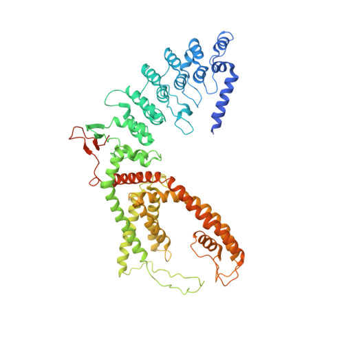
    Epithelial calcium channel TRPV6 plays vital roles in calcium homeostasis, and its dysregulation is implicated in multifactorial diseases, including cancers. Here, we study the molecular mechanism of selective nanomolar-affinity TRPV6 inhibition by (4-phenylcyclohexyl)piperazine derivatives (PCHPDs). We use x-ray crystallography and cryo-electron microscopy to solve the inhibitor-bound structures of TRPV6 and identify two types of inhibitor binding sites in the transmembrane region: (i) modulatory sites between the S1-S4 and pore domains normally occupied by lipids and (ii) the main site in the ion channel pore. Our structural data combined with mutagenesis, functional and computational approaches suggest that PCHPDs plug the open pore of TRPV6 and convert the channel into a nonconducting state, mimicking the action of calmodulin, which causes inactivation of TRPV6 channels under physiological conditions. This mechanism of inhibition explains the high selectivity and potency of PCHPDs and opens up unexplored avenues for the design of future-generation biomimetic drugs.


  • Organizational Affiliation

    Department of Nephrology and Hypertension and Department of Biomedical Research, University of Bern, Inselspital, Freiburgstrasse 15, CH-3010 Bern, Switzerland.


Macromolecules
Find similar proteins by:  (by identity cutoff)  |  3D Structure
Entity ID: 1
MoleculeChains Sequence LengthOrganismDetailsImage
Transient receptor potential cation channel subfamily V member 6672Rattus norvegicusMutation(s): 3 
Gene Names: Trpv6
Membrane Entity: Yes 
UniProt
Find proteins for Q9R186 (Rattus norvegicus)
Explore Q9R186 
Go to UniProtKB:  Q9R186
Entity Groups  
Sequence Clusters30% Identity50% Identity70% Identity90% Identity95% Identity100% Identity
UniProt GroupQ9R186
Sequence Annotations
Expand
  • Reference Sequence
Experimental Data & Validation

Experimental Data

  • Method: X-RAY DIFFRACTION
  • Resolution: 3.70 Å
  • R-Value Free: 0.327 
  • R-Value Work: 0.316 
  • R-Value Observed: 0.317 
  • Space Group: P 4 21 2
Unit Cell:
Length ( Å )Angle ( ˚ )
a = 146.228α = 90
b = 146.228β = 90
c = 114.648γ = 90
Software Package:
Software NamePurpose
PHENIXrefinement
XDSdata reduction
XDSdata scaling
PHENIXphasing

Structure Validation

View Full Validation Report


Ligand Structure Quality Assessment 


Entry History & Funding Information

Deposition Data


Funding OrganizationLocationGrant Number
National Institutes of Health/National Cancer Institute (NIH/NCI)United StatesR01 CA206573

Revision History  (Full details and data files)

  • Version 1.0: 2021-10-06
    Type: Initial release
  • Version 1.1: 2021-10-20
    Changes: Database references, Structure summary
  • Version 1.2: 2023-04-19
    Changes: Database references
  • Version 1.3: 2023-11-29
    Changes: Data collection, Refinement description