


Crystal structure of sulfotransferase Sult2A8 reveals the distinct mode of substrate binding for sulfonation at 7-hydroxyl group
Wang, K., Chan, Y.C., Lee, S.S.T., Au, S.W.N.To be published.
Experimental Data Snapshot
Starting Model: experimental
View more details
Entity ID: 1 | |||||
|---|---|---|---|---|---|
| Molecule | Chains | Sequence Length | Organism | Details | Image |
| Sulfotransferase | 284 | Mus musculus | Mutation(s): 0 Gene Names: Sult2a8, 2810007J24Rik EC: 2.8.2 (PDB Primary Data), 2.8.2.34 (UniProt) | 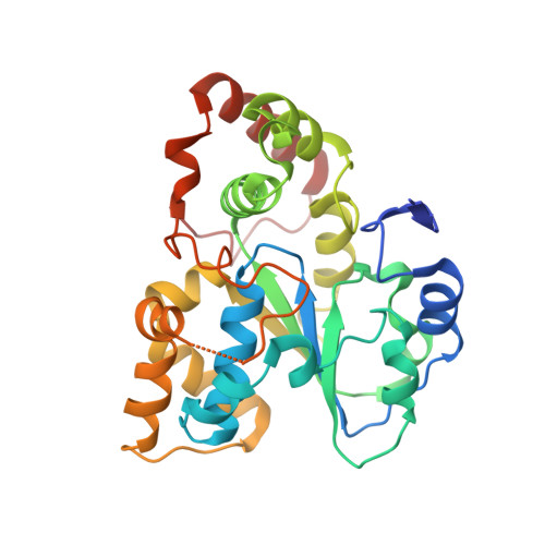 | |
UniProt & NIH Common Fund Data Resources | |||||
IMPC: MGI:1924221 | |||||
Entity Groups | |||||
| Sequence Clusters | 30% Identity50% Identity70% Identity90% Identity95% Identity100% Identity | ||||
| UniProt Group | Q8BGL3 | ||||
Sequence AnnotationsExpand | |||||
Reference Sequence | |||||
| Ligands 2 Unique | |||||
|---|---|---|---|---|---|
| ID | Chains | Name / Formula / InChI Key | 2D Diagram | 3D Interactions | |
| A3P Download:Ideal Coordinates CCD File | D [auth A], F [auth B] | ADENOSINE-3'-5'-DIPHOSPHATE C10 H15 N5 O10 P2 WHTCPDAXWFLDIH-KQYNXXCUSA-N |  | ||
| CHD (Subject of Investigation/LOI) Download:Ideal Coordinates CCD File | C [auth A], E [auth B] | CHOLIC ACID C24 H40 O5 BHQCQFFYRZLCQQ-OELDTZBJSA-N |  | ||
| Length ( Å ) | Angle ( ˚ ) |
|---|---|
| a = 93.87 | α = 90 |
| b = 93.87 | β = 90 |
| c = 71.3 | γ = 90 |
| Software Name | Purpose |
|---|---|
| PHENIX | refinement |
| CrystalClear | data collection |
| Coot | model building |
| SCALEPACK | data scaling |
| PHENIX | phasing |
| HKL-2000 | data reduction |


