7CZE

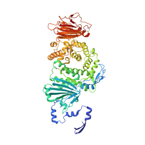
Crystal structure of Epstein-Barr virus (EBV) gHgL and in complex with the ligand binding domian (LBD) of EphA2


Experimental Data Snapshot

  • Method: X-RAY DIFFRACTION
  • Resolution: 3.00 Å
  • R-Value Free: 0.285 
  • R-Value Work: 0.257 
  • R-Value Observed: 0.259 

Starting Models: experimental
View more details

wwPDB Validation   3D Report Full Report


Ligand Structure Quality Assessment 


This is version 1.6 of the entry. See complete history


Literature

Molecular basis of EphA2 recognition by gHgL from gammaherpesviruses.

Su, C.Wu, L.Chai, Y.Qi, J.Tan, S.Gao, G.F.Song, H.Yan, J.

(2020) Nat Commun 11: 5964-5964

  • DOI: https://doi.org/10.1038/s41467-020-19617-9
  • Primary Citation of Related Structures:  
    7CZE, 7CZF

  • PubMed Abstract: 

    The human γ-herpesviruses Kaposi sarcoma associated herpesvirus (KSHV) and Epstein-Barr virus (EBV) are associated with many human malignancies. Viral glycoprotein H (gH) and glycoprotein L (gL) are crucial for the cell tropism by binding to specific receptors. Recently, EphA2 was identified as the specific entry receptor for both KSHV and EBV. Here, we characterized the crystal structures of KSHV gHgL or EBV gHgL in complex with the ligand binding domain (LBD) of EphA2. Both KSHV and EBV gHgL bind to the channel and peripheral regions of LBD primarily using gL. Extensive interactions with more contacts contribute to the higher affinity of KSHV gHgL to LBD than that of EBV gHgL. These binding characteristics were verified using cell-based fusion assays with mutations in key EphA2 residues. Our experiments suggest that multiple animal γ-herpesviruses could use EphA2 as an entry receptor, implying a potential threat to human health.


  • Organizational Affiliation

    College of Veterinary Medicine, China Agricultural University, Beijing, 100193, China.


Macromolecules
Find similar proteins by:  (by identity cutoff)  |  3D Structure
Entity ID: 1
MoleculeChains Sequence LengthOrganismDetailsImage
Envelope glycoprotein H
A, C, E, G
662Human herpesvirus 4 strain B95-8Mutation(s): 0 
Gene Names: gHBXLF2
UniProt
Find proteins for P03231 (Epstein-Barr virus (strain B95-8))
Explore P03231 
Go to UniProtKB:  P03231
Entity Groups  
Sequence Clusters30% Identity50% Identity70% Identity90% Identity95% Identity100% Identity
UniProt GroupP03231
Glycosylation
Glycosylation Sites: 2
Sequence Annotations
Expand
  • Reference Sequence
Find similar proteins by:  (by identity cutoff)  |  3D Structure
Entity ID: 2
MoleculeChains Sequence LengthOrganismDetailsImage
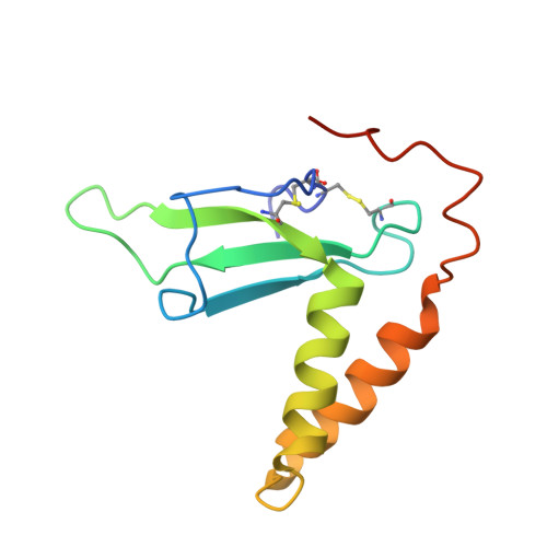
Envelope glycoprotein L
B, D, F, H
114Human herpesvirus 4 strain B95-8Mutation(s): 0 
Gene Names: gLBKRF2
UniProt
Find proteins for P03212 (Epstein-Barr virus (strain B95-8))
Explore P03212 
Go to UniProtKB:  P03212
Entity Groups  
Sequence Clusters30% Identity50% Identity70% Identity90% Identity95% Identity100% Identity
UniProt GroupP03212
Glycosylation
Glycosylation Sites: 2
Sequence Annotations
Expand
  • Reference Sequence
Find similar proteins by:  (by identity cutoff)  |  3D Structure
Entity ID: 3
MoleculeChains Sequence LengthOrganismDetailsImage
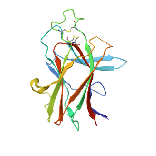
Ephrin type-A receptor 2
I, J, K, L
183Homo sapiensMutation(s): 0 
Gene Names: EPHA2ECK
EC: 2.7.10.1
UniProt & NIH Common Fund Data Resources
Find proteins for P29317 (Homo sapiens)
Explore P29317 
Go to UniProtKB:  P29317
PHAROS:  P29317
GTEx:  ENSG00000142627 
Entity Groups  
Sequence Clusters30% Identity50% Identity70% Identity90% Identity95% Identity100% Identity
UniProt GroupP29317
Sequence Annotations
Expand
  • Reference Sequence
Small Molecules
Ligands 1 Unique
IDChains Name / Formula / InChI Key2D Diagram3D Interactions
NAG (Subject of Investigation/LOI)
Query on NAG

Download Ideal Coordinates CCD File 
AA [auth H]
BA [auth H]
M [auth A]
N [auth A]
O [auth B]
AA [auth H],
BA [auth H],
M [auth A],
N [auth A],
O [auth B],
P [auth B],
Q [auth C],
R [auth C],
S [auth D],
T [auth D],
U [auth E],
V [auth E],
W [auth F],
X [auth F],
Y [auth G],
Z [auth G]
2-acetamido-2-deoxy-beta-D-glucopyranose
C8 H15 N O6
OVRNDRQMDRJTHS-FMDGEEDCSA-N
Experimental Data & Validation

Experimental Data

  • Method: X-RAY DIFFRACTION
  • Resolution: 3.00 Å
  • R-Value Free: 0.285 
  • R-Value Work: 0.257 
  • R-Value Observed: 0.259 
  • Space Group: P 1
Unit Cell:
Length ( Å )Angle ( ˚ )
a = 94.464α = 90.03
b = 113.806β = 90.167
c = 119.654γ = 89.888
Software Package:
Software NamePurpose
HKL-2000data scaling
PHENIXrefinement
HKL-2000data reduction
PHASERphasing

Structure Validation

View Full Validation Report


Ligand Structure Quality Assessment 


Entry History 

Deposition Data

Revision History  (Full details and data files)

  • Version 1.0: 2020-10-21
    Type: Initial release
  • Version 1.1: 2020-10-28
    Changes: Data collection
  • Version 1.2: 2020-12-02
    Changes: Database references
  • Version 1.3: 2020-12-09
    Changes: Database references
  • Version 1.4: 2021-01-13
    Changes: Other
  • Version 1.5: 2021-09-29
    Changes: Database references, Other
  • Version 1.6: 2023-11-29
    Changes: Data collection, Refinement description