7CZ2 | pdb_00007cz2

The complex structure of MSMEG_1954-ADP from Mycobacterium smegmatis


Experimental Data Snapshot

  • Method: X-RAY DIFFRACTION
  • Resolution: 1.80 Å
  • R-Value Free: 
    0.224 (Depositor), 0.231 (DCC) 
  • R-Value Work: 
    0.191 (Depositor), 0.200 (DCC) 
  • R-Value Observed: 
    0.193 (Depositor) 

Starting Model: experimental
View more details

wwPDB Validation 3D Report Full Report

Validation slider image for 7CZ2

Ligand Structure Quality Assessment 


This is version 1.2 of the entry. See complete history

Literature

Conformational Changes in a Macrolide Antibiotic Binding Protein From Mycobacterium smegmatis Upon ADP Binding.

Zhang, Q.Liu, X.Liu, H.Zhang, B.Yang, H.Mi, K.Guddat, L.W.Rao, Z.

(2021) Front Microbiol 12: 780954-780954

  • DOI: https://doi.org/10.3389/fmicb.2021.780954
  • Primary Citation Related Structures: 
    7CY2, 7CYR, 7CZ2

  • PubMed Abstract: 

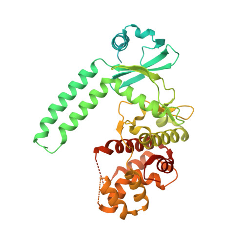
    Rv3197 (MABP-1), a non-canonical ABC protein in Mycobacterium tuberculosis , has ATPase activity and confers inducible resistance to the macrolide family of antibiotics. Here we have shown that MSMEG_1954, the homolog of Rv3197 in M. smegmatis , has a similar function of conferring macrolide resistance. Crystal structures of apo-MSMEG_1954 (form1 and form 2) and MSMEG_1954 in complex with ADP have been determined. These three structures show that MSMEG_1954 has at least two different conformations we identify as closed state (MSMEG_1954-form 1) and open state (MSMEG_1954-form 2 and MSMEG_1954-ADP). Structural superimposition shows that the MSMEG_1954-form 2 and MSMEG_1954-ADP complex have similar conformation to that observed for MABP-1 and MABP-1-erythromicin complex structure. However, the antibiotic binding pocket in MSMEG_1954-form 1 is completely blocked by the N-terminal accessory domain. When bound by ADP, the N-terminal accessory domain undergoes conformational change, which results in the open of the antibiotic binding pocket. Because of the degradation of N terminal accessory domain in MSMSG_1954-form 2, it is likely to represent a transitional state between MSMEG_1954-form 1 and MSMEG_1954-ADP complex structure.


  • Organizational Affiliation
    • State Key Laboratory of Medicinal Chemical Biology, Frontiers Science Center for Cell Responses, College of Life Sciences, Nankai University, Tianjin, China.

Macromolecule Content 

  • Total Structure Weight: 48.51 kDa 
  • Atom Count: 2,780 
  • Modeled Residue Count: 313 
  • Deposited Residue Count: 439 
  • Unique protein chains: 1

Macromolecules

Find similar proteins by:|  3D Structure
Entity ID: 1
MoleculeChains  Sequence LengthOrganismDetailsImage
ABC1 family protein439Mycolicibacterium smegmatis MC2 155Mutation(s): 0 
Gene Names: MSMEG_1954
UniProt
Find proteins for A0QTT2 (Mycolicibacterium smegmatis (strain ATCC 700084 / mc(2)155))
Explore A0QTT2 
Go to UniProtKB:  A0QTT2
Entity Groups
Sequence Clusters30% Identity50% Identity70% Identity90% Identity95% Identity100% Identity
UniProt GroupA0QTT2
Sequence Annotations
Expand
Reference Sequence

Experimental Data & Validation

Experimental Data

  • Method: X-RAY DIFFRACTION
  • Resolution: 1.80 Å
  • R-Value Free:  0.224 (Depositor), 0.231 (DCC) 
  • R-Value Work:  0.191 (Depositor), 0.200 (DCC) 
  • R-Value Observed: 0.193 (Depositor) 
Space Group: P 32 2 1
Unit Cell:
Length ( Å )Angle ( ˚ )
a = 83.65α = 90
b = 83.65β = 90
c = 100.76γ = 120
Software Package:
Software NamePurpose
REFMACrefinement
HKL-2000data scaling
PDB_EXTRACTdata extraction
HKL-2000data reduction
PHASERphasing

Structure Validation

View Full Validation Report


Ligand Structure Quality Assessment 


Entry History 

& Funding Information

Deposition Data


Funding OrganizationLocationGrant Number
National Natural Science Foundation of China (NSFC)China81520108019

Revision History  (Full details and data files)

  • Version 1.0: 2021-12-08
    Type: Initial release
  • Version 1.1: 2022-02-23
    Changes: Database references
  • Version 1.2: 2023-11-29
    Changes: Data collection, Refinement description