7CYU

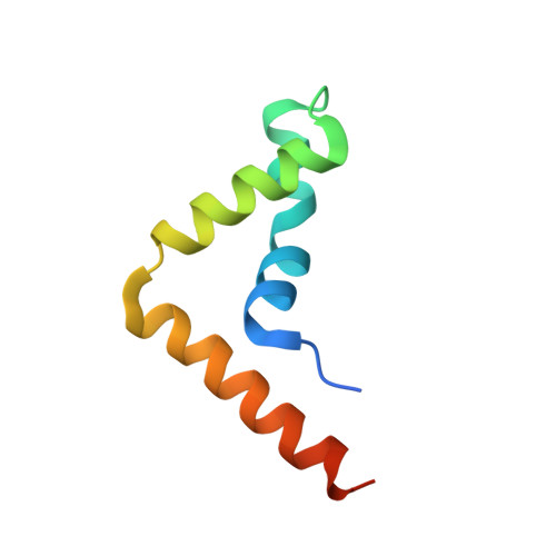
Crystal structure of human BAF57 HMG domain


Experimental Data Snapshot

  • Method: X-RAY DIFFRACTION
  • Resolution: 2.55 Å
  • R-Value Free: 0.275 
  • R-Value Work: 0.233 
  • R-Value Observed: 0.238 

Starting Model: experimental
View more details

wwPDB Validation   3D Report Full Report


This is version 1.2 of the entry. See complete history


Literature

Crystal structure of the HMG domain of human BAF57 and its interaction with four-way junction DNA.

Heo, Y.Park, J.H.Kim, J.Han, J.Yun, J.H.Lee, W.

(2020) Biochem Biophys Res Commun 533: 919-924

  • DOI: https://doi.org/10.1016/j.bbrc.2020.09.094
  • Primary Citation of Related Structures:  
    7CYU

  • PubMed Abstract: 

    The SWI/SNF chromatin remodeling complex plays important roles in gene regulation and it is classified as the SWI/SNF complex in yeast and BAF complex in vertebrates. BAF57, one of the subunits that forms the chromatin remodeling complex core, is well conserved in the BAF complex of vertebrates, which is replaced by bap111 in the Drosophila BAP complex and does not have a counterpart in the yeast SWI/SNF complex. This suggests that BAF57 is a key component of the chromatin remodeling complex in higher eukaryotes. BAF57 contains a HMG domain, which is widely distributed among various proteins and functions as a DNA binding motif. Most proteins with HMG domain bind to four-way junction (4WJ) DNA. Here, we report the crystal structure of the HMG domain of BAF57 (BAF57 HMG ) at a resolution of 2.55 Å. The structure consists of three α-helices and adopts an L-shaped form. The overall structure is stabilized by a hydrophobic core, which is formed by hydrophobic residues. The binding affinity between BAF57 HMG and 4WJ DNA is determined as a 295.83 ± 1.05 nM using a fluorescence quenching assay, and the structure revealed 4WJ DNA binding site of BAF57 HMG . Our data will serve structural basis in understanding the roles of BAF57 during chromatin remodeling process.


  • Organizational Affiliation

    Structural Biochemistry & Molecular Biophysics Laboratory, Department of Biochemistry, College of Life Sciences & Biotechnology, Yonsei University, Seoul, 120-749, South Korea.


Macromolecules
Find similar proteins by:  (by identity cutoff)  |  3D Structure
Entity ID: 1
MoleculeChains Sequence LengthOrganismDetailsImage
SWI/SNF-related matrix-associated actin-dependent regulator of chromatin subfamily E member 171Homo sapiensMutation(s): 0 
Gene Names: SMARCE1BAF57
UniProt & NIH Common Fund Data Resources
Find proteins for Q969G3 (Homo sapiens)
Explore Q969G3 
Go to UniProtKB:  Q969G3
PHAROS:  Q969G3
GTEx:  ENSG00000073584 
Entity Groups  
Sequence Clusters30% Identity50% Identity70% Identity90% Identity95% Identity100% Identity
UniProt GroupQ969G3
Sequence Annotations
Expand
  • Reference Sequence
Experimental Data & Validation

Experimental Data

  • Method: X-RAY DIFFRACTION
  • Resolution: 2.55 Å
  • R-Value Free: 0.275 
  • R-Value Work: 0.233 
  • R-Value Observed: 0.238 
  • Space Group: P 32 2 1
Unit Cell:
Length ( Å )Angle ( ˚ )
a = 58.873α = 90
b = 58.873β = 90
c = 50.263γ = 120
Software Package:
Software NamePurpose
PHENIXrefinement
HKL-2000data reduction
HKL-2000data scaling
PHENIXphasing

Structure Validation

View Full Validation Report


Entry History & Funding Information

Deposition Data


Funding OrganizationLocationGrant Number
National Research Foundation (NRF, Korea)Korea, Republic Of--

Revision History  (Full details and data files)

  • Version 1.0: 2020-10-14
    Type: Initial release
  • Version 1.1: 2020-12-30
    Changes: Database references
  • Version 1.2: 2023-11-29
    Changes: Data collection, Database references, Refinement description