Characterization of Enzymes Catalyzing the Formation of the Nonproteinogenic Amino Acid l-Dap in Capreomycin Biosynthesis.
Hsu, S.H., Zhang, S., Huang, S.C., Wu, T.K., Xu, Z., Chang, C.Y.(2021) Biochemistry 60: 77-84
- PubMed: 33356147 Search on PubMed
- DOI: https://doi.org/10.1021/acs.biochem.0c00808
- Primary Citation Related Structures:
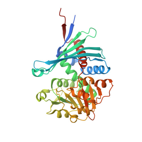
7CXS, 7CXU, 7CXV - PubMed Abstract:
Capreomycin (CMN) and viomycin (VIO) are nonribosomal peptide antituberculosis antibiotics, the structures of which contain four nonproteinogenic amino acids, including l-2,3-diaminopropionic acid (l-Dap), β-ureidodehydroalanine, l-capreomycidine, and β-lysine. Previous bioinformatics analysis suggested that CmnB/VioB and CmnK/VioK participate in the formation of l-Dap; however, the real substrates of these enzymes are yet to be confirmed. We herein show that starting from O -phospho-l-Ser (OPS) and l-Glu precursors, CmnB catalyzes the condensation reaction to generate a metabolite intermediate N -(1-amino-1-carboxyl-2-ethyl)glutamic acid (ACEGA), which undergoes NAD + -dependent oxidative hydrolysis by CmnK to generate l-Dap. Furthermore, the binding site of ACEGA and the catalytic mechanism of CmnK were elucidated with the assistance of three crystal structures, including those of apo-CmnK, the NAD + -CmnK complex, and CmnK in an alternative conformation. The CmnK-ACEGA docking model revealed that the glutamate α-hydrogen points toward the nicotinamide moiety. It provides evidence that the reaction is dependent on hydride transfer to form an imine intermediate, which is subsequently hydrolyzed by a water molecule to produce l-Dap. These findings modify the original proposed pathway and provide insights into l-Dap formation in the biosynthesis of other related natural products.
- Department of Biological Science and Technology, National Chiao Tung University, Hsinchu 30010, Taiwan, ROC.
Organizational Affiliation: