7CX5

Crystal structure of the DNA-binding domain of Bacillus subtilis CssR


Experimental Data Snapshot

  • Method: X-RAY DIFFRACTION
  • Resolution: 1.07 Å
  • R-Value Free: 0.204 
  • R-Value Work: 0.194 
  • R-Value Observed: 0.195 

wwPDB Validation   3D Report Full Report


This is version 1.1 of the entry. See complete history


Literature

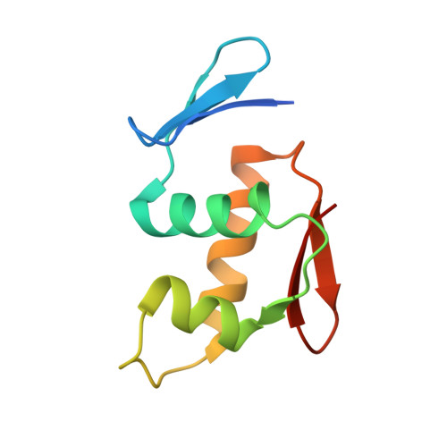
Crystal structure of the DNA-binding domain of Bacillus subtilis CssR.

Dahal, P.Kim, D.Y.Kwon, E.

(2021) Biochem Biophys Res Commun 555: 26-31

  • DOI: https://doi.org/10.1016/j.bbrc.2021.03.101
  • Primary Citation of Related Structures:  
    7CX5

  • PubMed Abstract: 

    Bacteria utilize two-component systems to regulate gene expression in response to changes in environmental stimuli. CssS/CssR, a two-component system in Bacillus subtilis, is responsible for overcoming envelope stresses caused by heat shock and secretion overload. During stress, the sensor component CssS is auto-phosphorylated and transfers the phosphoryl group to the response regulator CssR. Phosphorylated CssR then directly regulates the transcription of genes required to counteract the stress. Here, the crystal structure of the DNA-binding domain of CssR, determined at 1.07 Å resolution, is reported. The structure shows that the DNA-binding domain of CssR harbors a winged helix-turn-helix motif that is conserved in the OmpR/PhoB subfamily of response regulators. Based on the crystal structure, the dimeric architecture of the full-length CssR and its DNA-binding mode were suggested.


  • Organizational Affiliation

    College of Pharmacy, Yeungnam University, Gyeongsan, Gyeongbuk, 38541, Republic of Korea.


Macromolecules
Find similar proteins by:  (by identity cutoff)  |  3D Structure
Entity ID: 1
MoleculeChains Sequence LengthOrganismDetailsImage
Transcriptional regulatory protein CssR95Bacillus subtilis subsp. subtilis str. 168Mutation(s): 0 
Gene Names: cssRBSU33010
UniProt
Find proteins for O32192 (Bacillus subtilis (strain 168))
Explore O32192 
Go to UniProtKB:  O32192
Entity Groups  
Sequence Clusters30% Identity50% Identity70% Identity90% Identity95% Identity100% Identity
UniProt GroupO32192
Sequence Annotations
Expand
  • Reference Sequence
Experimental Data & Validation

Experimental Data

  • Method: X-RAY DIFFRACTION
  • Resolution: 1.07 Å
  • R-Value Free: 0.204 
  • R-Value Work: 0.194 
  • R-Value Observed: 0.195 
  • Space Group: P 21 21 21
Unit Cell:
Length ( Å )Angle ( ˚ )
a = 29.41α = 90
b = 52.74β = 90
c = 58.99γ = 90
Software Package:
Software NamePurpose
PHENIXrefinement
PDB_EXTRACTdata extraction
HKL-2000data reduction
HKL-2000data scaling
PHENIXphasing

Structure Validation

View Full Validation Report


Entry History 

Deposition Data

Revision History  (Full details and data files)

  • Version 1.0: 2021-04-21
    Type: Initial release
  • Version 1.1: 2024-03-27
    Changes: Data collection, Database references