7CVT

Crystal structure of the C85A/L194A/H234C mutant CLC-ec1 with Fab fragment


Experimental Data Snapshot

  • Method: X-RAY DIFFRACTION
  • Resolution: 2.90 Å
  • R-Value Free: 0.241 
  • R-Value Work: 0.201 
  • R-Value Observed: 0.203 

wwPDB Validation   3D Report Full Report


This is version 1.2 of the entry. See complete history


Literature

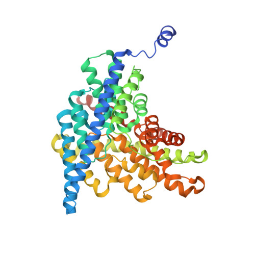
Altering CLC stoichiometry by reducing non-polar side-chains at the dimerization interface.

Mersch, K.Ozturk, T.N.Park, K.Lim, H.H.Robertson, J.L.

(2021) J Mol Biol 433: 166886-166886

  • DOI: https://doi.org/10.1016/j.jmb.2021.166886
  • Primary Citation of Related Structures:  
    7CVS, 7CVT

  • PubMed Abstract: 

    CLC-ec1 is a Cl - /H + antiporter that forms stable homodimers in lipid bilayers, with a free energy of -10.9 kcal/mol in 2:1 POPE/POPG lipid bilayers. The dimerization interface is formed by four transmembrane helices: H, I, P and Q, that are lined by non-polar side-chains that come in close contact, yet it is unclear as to whether their interactions drive dimerization. To investigate whether non-polar side-chains are required for dimer assembly, we designed a series of constructs where side-chain packing in the dimer state is significantly reduced by making 4-5 alanine substitutions along each helix (H-ala, I-ala, P-ala, Q-ala). All constructs are functional and three purify as stable dimers in detergent micelles despite the removal of significant side-chain interactions. On the other hand, H-ala shows the unique behavior of purifying as a mixture of monomers and dimers, followed by a rapid and complete conversion to monomers. In lipid bilayers, all four constructs are monomeric as examined by single-molecule photobleaching analysis. Further study of the H-helix shows that the single mutation L194A is sufficient to yield monomeric CLC-ec1 in detergent micelles and lipid bilayers. X-ray crystal structures of L194A reveal the protein re-assembles to form dimers, with a structure that is identical to wild-type. Altogether, these results demonstrate that non-polar membrane embedded side-chains play an important role in defining dimer stability, but the stoichiometry is highly contextual to the solvent environment. Furthermore, we discovered that L194 is a molecular hot-spot for defining dimerization of CLC-ec1.


  • Organizational Affiliation

    Department of Biochemistry and Molecular Biophysics, Washington University School of Medicine in St. Louis, St. Louis, MO, USA; Department of Molecular Physiology and Biophysics, The University of Iowa, Iowa City, IA, USA.


Macromolecules
Find similar proteins by:  (by identity cutoff)  |  3D Structure
Entity ID: 1
MoleculeChains Sequence LengthOrganismDetailsImage
H(+)/Cl(-) exchange transporter ClcA
A, B
473Escherichia coli MS 198-1Mutation(s): 3 
Gene Names: clcAeriCHMPREF9552_04837
Membrane Entity: Yes 
UniProt
Find proteins for P37019 (Escherichia coli (strain K12))
Explore P37019 
Go to UniProtKB:  P37019
Entity Groups  
Sequence Clusters30% Identity50% Identity70% Identity90% Identity95% Identity100% Identity
UniProt GroupP37019
Sequence Annotations
Expand
  • Reference Sequence
Find similar proteins by:  (by identity cutoff)  |  3D Structure
Entity ID: 2
MoleculeChains Sequence LengthOrganismDetailsImage
antibody Fab fragment heavy chain
C, E
222Mus musculusMutation(s): 0 
Entity Groups  
Sequence Clusters30% Identity50% Identity70% Identity90% Identity95% Identity100% Identity
Sequence Annotations
Expand
  • Reference Sequence
Find similar proteins by:  (by identity cutoff)  |  3D Structure
Entity ID: 3
MoleculeChains Sequence LengthOrganismDetailsImage
antibody Fab fragment light chain
D, F
211Mus musculusMutation(s): 0 
Entity Groups  
Sequence Clusters30% Identity50% Identity70% Identity90% Identity95% Identity100% Identity
Sequence Annotations
Expand
  • Reference Sequence
Experimental Data & Validation

Experimental Data

  • Method: X-RAY DIFFRACTION
  • Resolution: 2.90 Å
  • R-Value Free: 0.241 
  • R-Value Work: 0.201 
  • R-Value Observed: 0.203 
  • Space Group: C 1 2 1
Unit Cell:
Length ( Å )Angle ( ˚ )
a = 233.034α = 90
b = 98.305β = 131.99
c = 172.434γ = 90
Software Package:
Software NamePurpose
PHENIXrefinement
HKL-2000data reduction
HKL-2000data scaling
PHASERphasing

Structure Validation

View Full Validation Report


Entry History & Funding Information

Deposition Data


Funding OrganizationLocationGrant Number
Ministry of Science, ICT and Future Planning (MSIP)Korea, Republic Of20-BR-01-05

Revision History  (Full details and data files)

  • Version 1.0: 2021-09-01
    Type: Initial release
  • Version 1.1: 2022-03-16
    Changes: Database references
  • Version 1.2: 2023-11-29
    Changes: Data collection, Refinement description