7CUC

Crystal Structure of Urate Oxidase from Bacillus sp. TB-90 in the absence from Chloride Anion at 1.44 A resolution


Experimental Data Snapshot

  • Method: X-RAY DIFFRACTION
  • Resolution: 1.44 Å
  • R-Value Free: 0.208 
  • R-Value Work: 0.175 
  • R-Value Observed: 0.177 

wwPDB Validation   3D Report Full Report


Ligand Structure Quality Assessment 


This is version 1.2 of the entry. See complete history


Literature

Identification of quasi-stable water molecules near the Thr73-Lys13 catalytic diad of Bacillus sp. TB-90 urate oxidase by X-ray crystallography with controlled humidity.

Hibi, T.Itoh, T.

(2021) J Biochem 169: 15-23

  • DOI: https://doi.org/10.1093/jb/mvaa114
  • Primary Citation of Related Structures:  
    7CUC, 7CUF, 7CUG

  • PubMed Abstract: 

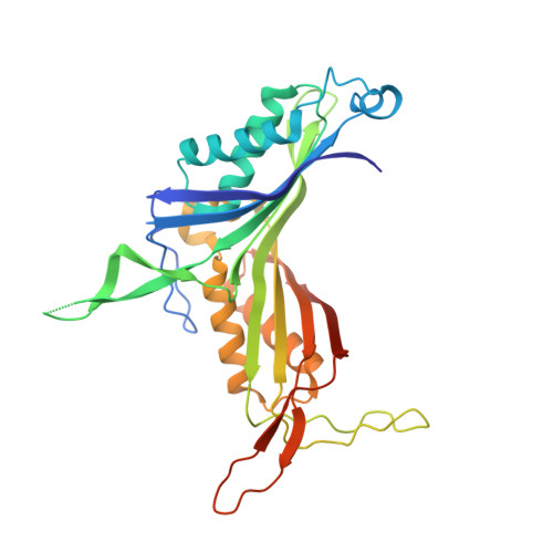
    Urate oxidases (UOs) catalyze the cofactor-independent oxidation of uric acid, and an extensive water network in the active site has been suggested to play an essential role in the catalysis. For our present analysis of the structure and function of the water network, the crystal qualities of Bacillus sp. TB-90 urate oxidase were improved by controlled dehydration using the humid air and glue-coating method. After the dehydration, the P21212 crystals were transformed into the I222 space group, leading to an extension of the maximum resolution to 1.42 Å. The dehydration of the crystals revealed a significant change in the five-water-molecules' binding mode in the vicinity of the catalytic diad, indicating that these molecules are quasi-stable. The pH profile analysis of log(kcat) gave two pKa values: pKa1 at 6.07 ± 0.07 and pKa2 at 7.98 ± 0.13. The site-directed mutagenesis of K13, T73 and N276 involved in the formation of the active-site water network revealed that the activities of these mutant variants were significantly reduced. These structural and kinetic data suggest that the five quasi-stable water molecules play an essential role in the catalysis of the cofactor-independent urate oxidation by reducing the energy penalty for the substrate-binding or an on-off switching for the proton-relay rectification.


  • Organizational Affiliation

    Department of Bioscience and Biotechnology, Fukui Prefectural University, 4-1-1 Matsuoka-Kenjojima, Eiheiji, Yoshida, Fukui 910-1195, Japan.


Macromolecules
Find similar proteins by:  (by identity cutoff)  |  3D Structure
Entity ID: 1
MoleculeChains Sequence LengthOrganismDetailsImage
Uric acid degradation bifunctional protein
A, B
313Bacillus sp. TB-90Mutation(s): 0 
Gene Names: uao
EC: 1.7.3.3
UniProt
Find proteins for Q45697 (Bacillus sp. (strain TB-90))
Explore Q45697 
Go to UniProtKB:  Q45697
Entity Groups  
Sequence Clusters30% Identity50% Identity70% Identity90% Identity95% Identity100% Identity
UniProt GroupQ45697
Sequence Annotations
Expand
  • Reference Sequence
Small Molecules
Ligands 5 Unique
IDChains Name / Formula / InChI Key2D Diagram3D Interactions
AZA (Subject of Investigation/LOI)
Query on AZA

Download Ideal Coordinates CCD File 
C [auth A],
L [auth B]
8-AZAXANTHINE
C4 H3 N5 O2
KVGVQTOQSNJTJI-UHFFFAOYSA-N
SO4
Query on SO4

Download Ideal Coordinates CCD File 
N [auth B]SULFATE ION
O4 S
QAOWNCQODCNURD-UHFFFAOYSA-L
MXE
Query on MXE

Download Ideal Coordinates CCD File 
F [auth A]
G [auth A]
H [auth A]
I [auth A]
J [auth B]
F [auth A],
G [auth A],
H [auth A],
I [auth A],
J [auth B],
K [auth B]
2-METHOXYETHANOL
C3 H8 O2
XNWFRZJHXBZDAG-UHFFFAOYSA-N
OXY
Query on OXY

Download Ideal Coordinates CCD File 
D [auth A],
M [auth B]
OXYGEN MOLECULE
O2
MYMOFIZGZYHOMD-UHFFFAOYSA-N
NA
Query on NA

Download Ideal Coordinates CCD File 
E [auth A],
O [auth B]
SODIUM ION
Na
FKNQFGJONOIPTF-UHFFFAOYSA-N
Experimental Data & Validation

Experimental Data

  • Method: X-RAY DIFFRACTION
  • Resolution: 1.44 Å
  • R-Value Free: 0.208 
  • R-Value Work: 0.175 
  • R-Value Observed: 0.177 
  • Space Group: I 2 2 2
Unit Cell:
Length ( Å )Angle ( ˚ )
a = 71.618α = 90
b = 133.836β = 90
c = 145.363γ = 90
Software Package:
Software NamePurpose
PHASERphasing
PHENIXrefinement
XDSdata processing
XDSdata scaling

Structure Validation

View Full Validation Report


Ligand Structure Quality Assessment 


Entry History 

Deposition Data

Revision History  (Full details and data files)

  • Version 1.0: 2020-11-25
    Type: Initial release
  • Version 1.1: 2021-02-17
    Changes: Database references
  • Version 1.2: 2023-11-29
    Changes: Data collection, Database references, Refinement description