7CPH

Crystal structure of tRNA adenosine deaminase from Bacillus subtilis


Experimental Data Snapshot

  • Method: X-RAY DIFFRACTION
  • Resolution: 2.30 Å
  • R-Value Free: 0.240 
  • R-Value Work: 0.177 
  • R-Value Observed: 0.181 

wwPDB Validation   3D Report Full Report


This is version 1.2 of the entry. See complete history


Literature

Deciphering protein microenvironment by using a cysteine specific switch-ON fluorescent probe.

Mariam, J.Hoskere Ashoka, A.Gaded, V.Ali, F.Malvi, H.Das, A.Anand, R.

(2021) Org Biomol Chem 19: 5161-5168

  • DOI: https://doi.org/10.1039/d1ob00698c
  • Primary Citation of Related Structures:  
    7CPH

  • PubMed Abstract: 

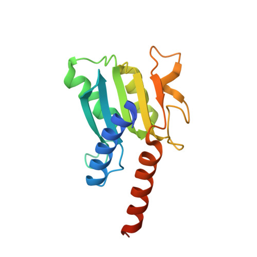
    Fluorescent probes provide an unparalleled opportunity to visualize and quantify dynamic events. Here, we employ a medium-size, cysteine specific coumarin based switch-ON fluorescent probe 'L' to track protein unfolding profiles and accessibility of cysteine residues in proteins. It was established that 'L' is highly selective and exhibits no artifact due to interaction with other bystander species. 'L' is able to gauge subtle changes in protein microenvironment and proved to be effective in delineating early unfolding events that are difficult to otherwise discern by classic techniques such as circular dichroism. By solving the X-ray structure of TadA and probing the temperature dependent fluorescence-ON response with native TadA and its cysteine mutants, it was revealed that unfolding occurs in a stage-wise manner and the regions that are functionally important form compact sub-domains and unfold at later stages. Our results assert that probe 'L' serves as an efficient tool to monitor subtle changes in protein structure and can be employed as a generic dye to study processes such as protein unfolding.


  • Organizational Affiliation

    Department of Chemistry, IIT Bombay, Mumbai-400076, India. ruchi@chem.iitb.ac.in.


Macromolecules
Find similar proteins by:  (by identity cutoff)  |  3D Structure
Entity ID: 1
MoleculeChains Sequence LengthOrganismDetailsImage
tRNA-specific adenosine deaminase
A, B
167Bacillus subtilis subsp. subtilis str. 168Mutation(s): 0 
Gene Names: tadAyaaJBSU00180
EC: 3.5.4.33
UniProt
Find proteins for P21335 (Bacillus subtilis (strain 168))
Explore P21335 
Go to UniProtKB:  P21335
Entity Groups  
Sequence Clusters30% Identity50% Identity70% Identity90% Identity95% Identity100% Identity
UniProt GroupP21335
Sequence Annotations
Expand
  • Reference Sequence
Experimental Data & Validation

Experimental Data

  • Method: X-RAY DIFFRACTION
  • Resolution: 2.30 Å
  • R-Value Free: 0.240 
  • R-Value Work: 0.177 
  • R-Value Observed: 0.181 
  • Space Group: P 21 21 21
Unit Cell:
Length ( Å )Angle ( ˚ )
a = 38.53α = 90
b = 41.25β = 90
c = 202.08γ = 90
Software Package:
Software NamePurpose
PHENIXrefinement
REFMACrefinement
iMOSFLMdata reduction
SCALAdata scaling
MOLREPphasing

Structure Validation

View Full Validation Report


Entry History & Funding Information

Deposition Data


Funding OrganizationLocationGrant Number
Other governmentIndiaEMR/2015/ 002121

Revision History  (Full details and data files)

  • Version 1.0: 2021-06-02
    Type: Initial release
  • Version 1.1: 2021-06-30
    Changes: Database references
  • Version 1.2: 2023-11-29
    Changes: Data collection, Database references, Refinement description